Texture Analysis of Torn Rotator Cuff on Preoperative Magnetic Resonance Arthrography as a Predictor of Postoperative Tendon Status
- Affiliations
-
- 1Department of Radiology, SMG-SNU Boramae Medical Center, Seoul National University College of Medicine, Seoul 07061, Korea.
- 2Department of Radiology, Seoul National University Bundang Hospital, Seongnam 13620, Korea. netty0523@gmail.com
- 3Department of Radiology, Chung-Ang University Hospital, Seoul 06973, Korea.
- 4Division of Biomedical Engineering, Hankuk University of Foreign Studies, Yongin 17035, Korea.
- KMID: 2427238
- DOI: http://doi.org/10.3348/kjr.2017.18.4.691
Abstract
OBJECTIVE
To evaluate texture data of the torn supraspinatus tendon (SST) on preoperative T2-weighted magnetic resonance arthrography (MRA) using the gray-level co-occurrence matrix (GLCM) for prediction of post-operative tendon state.
MATERIALS AND METHODS
Fifty patients who underwent arthroscopic rotator cuff repair for full-thickness tears of the SST were included in this retrospective study. Based on 1-year follow-up, magnetic resonance imaging showed that 30 patients had intact SSTs, and 20 had rotator cuff retears. Using GLCM, two radiologists measured independantly the highest signal intensity area of the distal end of the torn SST on preoperative T2-weighted MRA, which were compared between two groups.The relationships with other well-known prognostic factors, including age, tear size (anteroposterior dimension), retraction size (mediolateral tear length), grade of fatty degeneration of the SST and infraspinatus tendon, and arthroscopic fixation technique (single or double row), also were evaluated.
RESULTS
Of all the GLCM features, the retear group showed significantly higher entropy (p < 0.001 and p = 0.001), variance (p = 0.030 and 0.011), and contrast (p = 0.033 and 0.012), but lower angular second moment (p < 0.001 and p = 0.002) and inverse difference moment (p = 0.027 and 0.027), as well as larger tear size (p = 0.001) and retraction size (p = 0.002) than the intact group. Retraction size (odds ratio [OR] = 3.053) and entropy (OR = 17.095) were significant predictors.
CONCLUSION
Texture analysis of torn SSTs on preoperative T2-weighted MRA using the GLCM may be helpful to predict postoperative tendon state after rotator cuff repair.
Keyword
MeSH Terms
Figure
-
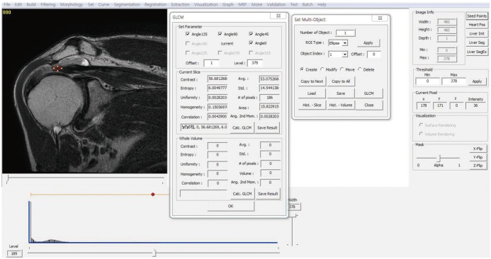
Fig. 1 GLCM program.Texture analysis of T2-weighted coronal images using gray-level co-occurrence matrix software package. GLCM = graylevel co-occurrence matrix
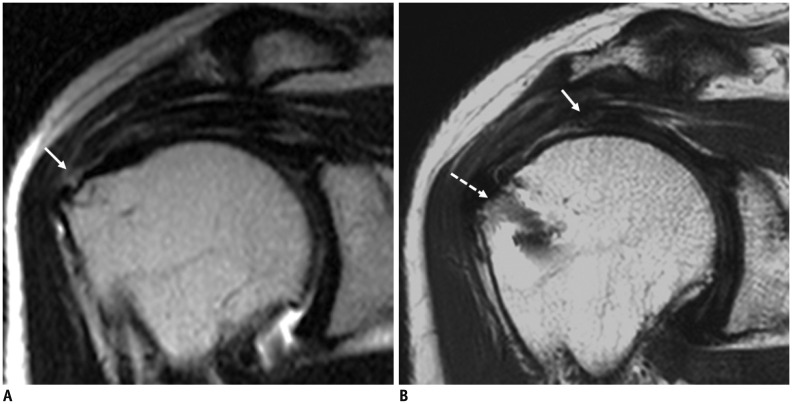
Fig. 2 70-year-old female with retear shows high entropy value measured by GLCM.A. Small-sized rim-rent tear of supraspinatus tendon is identified at greater tuberosity in 70-year-old woman on T2-weighted coronal image of right shoulder MRI (arrow), which has high entropy value measured by GLCM. B. One year later, she has retear of supraspinatus tendon (arrow) with large retraction from anchor of rotator cuff repair (dashed arrow) on postoperative MRI. GLCM = gray-level co-occurrence matrix
Cited by 2 articles
-
Age of Data in Contemporary Research Articles Published in Representative General Radiology Journals
Ji Hun Kang, Dong Hwan Kim, Seong Ho Park, Jung Hwan Baek
Korean J Radiol. 2018;19(6):1172-1178. doi: 10.3348/kjr.2018.19.6.1172.Evaluation of the Subscapularis Tendon Tears on 3T Magnetic Resonance Arthrography: Comparison of Diagnostic Performance of T1-Weighted Spectral Presaturation with Inversion-Recovery and T2-Weighted Turbo Spin-Echo Sequences
Hoseok Lee, Joong Mo Ahn, Yusuhn Kang, Joo Han Oh, Eugene Lee, Joon Woo Lee, Heung Sik Kang
Korean J Radiol. 2018;19(2):320-327. doi: 10.3348/kjr.2018.19.2.320.
Reference
-
1. Boileau P, Brassart N, Watkinson DJ, Carles M, Hatzidakis AM, Krishnan SG. Arthroscopic repair of full-thickness tears of the supraspinatus: does the tendon really heal? J Bone Joint Surg Am. 2005; 87:1229–1240. PMID: 15930531.
Article2. Huijsmans PE, Pritchard MP, Berghs BM, van Rooyen KS, Wallace AL, de Beer JF. Arthroscopic rotator cuff repair with double-row fixation. J Bone Joint Surg Am. 2007; 89:1248–1257. PMID: 17545428.
Article3. Le BT, Wu XL, Lam PH, Murrell GA. Factors predicting rotator cuff retears: an analysis of 1000 consecutive rotator cuff repairs. Am J Sports Med. 2014; 42:1134–1142. PMID: 24748610.4. Jost B, Zumstein M, Pfirrmann CW, Gerber C. Long-term outcome after structural failure of rotator cuff repairs. J Bone Joint Surg Am. 2006; 88:472–479. PMID: 16510810.
Article5. Harryman DT 2nd, Mack LA, Wang KY, Jackins SE, Richardson ML, Matsen FA 3rd. Repairs of the rotator cuff. Correlation of functional results with integrity of the cuff. J Bone Joint Surg Am. 1991; 73:982–989. PMID: 1874784.
Article6. Denard PJ, Burkhart SS. Arthroscopic revision rotator cuff repair. J Am Acad Orthop Surg. 2011; 19:657–666. PMID: 22052642.
Article7. Jost B, Pfirrmann CW, Gerber C, Switzerland Z. Clinical outcome after structural failure of rotator cuff repairs. J Bone Joint Surg Am. 2000; 82:304–314. PMID: 10724223.
Article8. Gladstone JN, Bishop JY, Lo IK, Flatow EL. Fatty infiltration and atrophy of the rotator cuff do not improve after rotator cuff repair and correlate with poor functional outcome. Am J Sports Med. 2007; 35:719–728. PMID: 17337727.
Article9. Nho SJ, Brown BS, Lyman S, Adler RS, Altchek DW, MacGillivray JD. Prospective analysis of arthroscopic rotator cuff repair: prognostic factors affecting clinical and ultrasound outcome. J Shoulder Elbow Surg. 2009; 18:13–20. PMID: 18799326.
Article10. Oh JH, Kim SH, Kang JY, Oh CH, Gong HS. Effect of age on functional and structural outcome after rotator cuff repair. Am J Sports Med. 2010; 38:672–678. PMID: 20357401.
Article11. Chung SW, Oh JH, Gong HS, Kim JY, Kim SH. Factors affecting rotator cuff healing after arthroscopic repair: osteoporosis as one of the independent risk factors. Am J Sports Med. 2011; 39:2099–2107. PMID: 21813440.12. Wu XL, Briggs L, Murrell GA. Intraoperative determinants of rotator cuff repair integrity: an analysis of 500 consecutive repairs. Am J Sports Med. 2012; 40:2771–2776. PMID: 23104609.13. Maqdes A, Abarca J, Moraiti C, Boughebri O, Dib C, Leclère FM, et al. Does preoperative subscapularis fatty muscle infiltration really matter in anterosuperior rotator cuff tears repair outcomes? A prospective multicentric study. Orthop Traumatol Surg Res. 2014; 100:485–488. PMID: 24947497.
Article14. Davnall F, Yip CS, Ljungqvist G, Selmi M, Ng F, Sanghera B, et al. Assessment of tumor heterogeneity: an emerging imaging tool for clinical practice? Insights Imaging. 2012; 3:573–589. PMID: 23093486.
Article15. Yun BL, Cho N, Li M, Jang MH, Park SY, Kang HC, et al. Intratumoral heterogeneity of breast cancer xenograft models: texture analysis of diffusion-weighted MR imaging. Korean J Radiol. 2014; 15:591–604. PMID: 25246820.
Article16. Loizou CP, Petroudi S, Seimenis I, Pantziaris M, Pattichis CS. Quantitative texture analysis of brain white matter lesions derived from T2-weighted MR images in MS patients with clinically isolated syndrome. J Neuroradiol. 2015; 42:99–114. PMID: 24970463.
Article17. Raja JV, Khan M, Ramachandra VK, Al-Kadi O. Texture analysis of CT images in the characterization of oral cancers involving buccal mucosa. Dentomaxillofac Radiol. 2012; 41:475–480. PMID: 22241875.
Article18. Haralick RM, Shanmugam K. Textural features for image classification. IEEE Trans Syst Man Cybern Syst. 1973; 3:610–621.
Article19. Sugaya H, Maeda K, Matsuki K, Moriishi J. Functional and structural outcome after arthroscopic full-thickness rotator cuff repair: single-row versus dual-row fixation. Arthroscopy. 2005; 21:1307–1316. PMID: 16325080.
Article20. Gebejes A, Huertas R. Texture characterization based on grey-level co-occurrence matrix. Conference of Informatics and Management Sciences. Zilina: EDIS-Publishing Institution of the University of Zilina;2013. p. 375–378.21. Mohanaiah P, Sathyanarayana P, GuruKumar L. Image texture feature extraction using GLCM approach. Int J Sci Res Publ. 2013; 3:1–5.22. Goutallier D, Postel JM, Bernageau J, Lavau L, Voisin MC. Fatty muscle degeneration in cuff ruptures. Pre- and postoperative evaluation by CT scan. Clin Orthop Relat Res. 1994; (304):78–83. PMID: 8020238.23. Mazzocca AD, Millett PJ, Guanche CA, Santangelo SA, Arciero RA. Arthroscopic single-row versus double-row suture anchor rotator cuff repair. Am J Sports Med. 2005; 33:1861–1868. PMID: 16210578.
Article24. Shen C, Tang ZH, Hu JZ, Zou GY, Xiao RC. Incidence of retear with double-row versus single-row rotator cuff repair. Orthopedics. 2014; 37:e1006–e1013. PMID: 25361362.
Article25. Cummins CA, Murrell GA. Mode of failure for rotator cuff repair with suture anchors identified at revision surgery. J Shoulder Elbow Surg. 2003; 12:128–133. PMID: 12700563.
Article26. Yip C, Landau D, Kozarski R, Ganeshan B, Thomas R, Michaelidou A, et al. Primary esophageal cancer: heterogeneity as potential prognostic biomarker in patients treated with definitive chemotherapy and radiation therapy. Radiology. 2014; 270:141–148. PMID: 23985274.
Article27. Zhang J, Tong L, Wang L, Li N. Texture analysis of multiple sclerosis: a comparative study. Magn Reson Imaging. 2008; 26:1160–1166. PMID: 18513908.
Article28. Riley GP, Harrall RL, Constant CR, Chard MD, Cawston TE, Hazleman BL. Tendon degeneration and chronic shoulder pain: changes in the collagen composition of the human rotator cuff tendons in rotator cuff tendinitis. Ann Rheum Dis. 1994; 53:359–366. PMID: 8037494.
Article29. Saridakis P, Jones G. Outcomes of single-row and double-row arthroscopic rotator cuff repair: a systematic review. J Bone Joint Surg Am. 2010; 92:732–742. PMID: 20194334.
Article30. Nozaki T, Tasaki A, Horiuchi S, Ochi J, Starkey J, Hara T, et al. Predicting Retear after repair of full-thickness rotator cuff tear: two-point dixon MR imaging quantification of fatty muscle degeneration-initial experience with 1-year follow-up. Radiology. 2016; 280:500–509. PMID: 26937710.
Article31. Lee JE, Park JS, Ryu KN, Rhee YG, Yoon SH, Park SY, et al. Repaired supraspinatus tendons in clinically improving patients: early postoperative findings and interval changes on MRI. Korean J Radiol. 2015; 16:363–371. PMID: 25741199.
Article32. Sugaya H, Maeda K, Matsuki K, Moriishi J. Repair integrity and functional outcome after arthroscopic double-row rotator cuff repair. A prospective outcome study. J Bone Joint Surg Am. 2007; 89:953–960. PMID: 17473131.
- Full Text Links
-
- Actions
-
Cited
- CITED
-
- Close
- Share
-
- Similar articles
-
- Arthrographic Pitfalls in the Diagnosis of Full-Thickness Tears of the Rotator Cuff: A Case Report
- Characteristics of Magnetic Resonance Arthrography Findings in Traumatic Posterosuperior Rotator Cuff Tears
- Postoperative Ultrasound Findings of the Rotator Cuff Tendon after Arthroscopic Repair of a Rotator Cuff Tear
- Factors Influencing the Restoration of Acromiohumeral Distance of Immediate Postoperative Period in Patients Who Have Rotator Cuff Repair Surgery with Large-to-Massive Rotator Cuff Tears
- Preoperative Three Dimensional Ultrasonographic Evaluation of the Rotator Cuff Tear
