Comparison of two techniques for transpharyngeal endoscopic auditory tube diverticulotomy in the horse
- Affiliations
-
- 1Department of Clinical Sciences, Colorado State University, Fort Collins, CO 80523, USA. Eileen.Hackett@colostate.edu
- 2Department of Biomedical Sciences, Colorado State University, Fort Collins, CO 80523, USA.
- KMID: 2427032
- DOI: http://doi.org/10.4142/jvs.2018.19.6.835
Abstract
- Auditory tube diverticula, also known as guttural pouches, are naturally occurring dilations of the auditory tube in horses that communicate with the nasopharynx through a small ostium. Infection and select other conditions can result in inflammation and narrowing of the nasopharyngeal ostium, which prevents drainage of fluid or egress of air and can lead to persistent infection or guttural pouch tympany. Auditory tube diverticulotomy allows continuous egress from the auditory tube diverticula and is a feature of disease treatment in horses, in which medical treatment alone is not successful. Transpharyngeal endoscopic auditory tube diverticulotomy was performed using a diode laser either at a single dorsal pharyngeal recess location or bilaterally caudal to the nasopharyngeal ostium in 10 horse head specimens. Both methods resulted in clear communication between the nasopharynx and auditory tube diverticula. Diverticulotomy performed in the dorsal pharyngeal recess required less laser energy and activation time and had a shorter surgical duration than diverticulotomy performed caudal to the nasopharyngeal ostium. Further study related to the clinical application of both techniques is warranted.
MeSH Terms
Figure
-

Fig. 1 Illustration of the equine nasopharynx. Incision areas for auditory tube diverticulotomy within the dorsal pharyngeal recess (DR) and caudal to the nasopharyngeal ostium (CNO) have been indicated by a red rectangle bordering the DR site and blue triangles bordering the CNO sites.

Fig. 2 Computed tomographic images (transverse plane [A] and dorsal plane [B]; 2 mm standard window) of a horse head specimen following auditory tube diverticulotomy within the dorsal pharyngeal recess. Note communication between the nasopharynx and both left and right auditory tube diverticula (white arrows). The median septum is visible (black arrow).
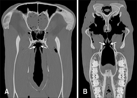
Fig. 3 Computed tomographic image (transverse plane [A] and dorsal plane [B]; 2 mm standard window) of a horse head specimen following auditory tube diverticulotomy caudal to the nasopharyngeal ostium. Note communication between the nasopharynx and both left and right auditory tube diverticula (white arrows). The median septum is visible (black arrow).
Reference
-
1. Adkins AR, Yovich JV, Colbourne CM. Nonsurgical treatment of chondroids of the guttural pouch in a horse. Aust Vet J. 1997; 75:332–333.
Article2. Baptiste KE, Naylor JM, Bailey J, Barber EM, Post K, Thornhill J. A function for guttural pouches in the horse. Nature. 2000; 403:382–383.
Article3. Cramp PA, Prange T, Nickels FA. Standing equine surgery of the upper respiratory tract. Vet Clin North Am Equine Pract. 2014; 30:111–141.
Article4. Edwards GB, Greet T. Disorders of the guttural pouches (auditory tube diverticuli). In : McGorum BC, Dixon PM, Robinson NE, Schumacher J, editors. Equine Respiratory Medicine and Surgery. Edinburgh: Elsevier Saunders;2007. p. 419–436.5. Freeman DE, Hardy J. Guttural pouch. In : Auer JA, Stick JA, editors. Equine Surgery. 4th ed. St. Louis: Elsevier/Saunders;2012. p. 623–642.6. Gehlen H, Ohnesorge B. Laser fenestration of the mesial septum for treatment of guttural pouch chondroids in a pony. Vet Surg. 2005; 34:383–386.
Article7. Hawkins JF, Frank N, Sojka JE, Levy M. Fistulation of the auditory tube diverticulum (guttural pouch) with a neodymium:yttrium-aluminum-garnet laser for treatment of chronic empyema in two horses. J Am Vet Med Assoc. 2001; 218:405–407.
Article8. Hinchcliffe R, Pye A. Variations in the middle ear of the Mammalia. J Zool. 1969; 157:277–288.
Article9. Judy CE, Chaffin MK, Cohen ND. Empyema of the guttural pouch (auditory tube diverticulum) in horses: 91 cases (1977-1997). J Am Vet Med Assoc. 1999; 215:1666–1670.10. Knight AP, Voss JL, McChesney AE, Bigbee HG. Experimentally-induced Streptococcus equi infection in horses with resultant guttural pouch empyema. Vet Med Small Anim Clin. 1975; 70:1194–1199.11. Krebs W, Schmotzer WB. Laser fenestrated salpingopharyngeal fistulas for treatment of bilateral guttural pouch tympany in a foal. Equine Vet Educ. 2007; 19:419–423.
Article12. Schambourg MA, Marcoux M, Céleste C. Salpingoscopy for the treatment of recurrent guttural pouch tympany in a filly. Equine Vet Educ. 2006; 18:231–234.
Article13. Seahorn TL, Schumacher J. Nonsurgical removal of chondroid masses from the guttural pouches of two horses. J Am Vet Med Assoc. 1991; 199:368–369.14. Sullin KE. Standing endoscopic application of cutting current for equine upper respiratory surgery. Equine Pract. 1994; 16:23–26.15. Tate LP. Application of lasers in equine upper respiratory surgery. Vet Clin North Am Equine Pract. 1991; 7:165–195.
Article16. Tate LP Jr, Blikslager AT, Little ED. Transendoscopic laser treatment of guttural pouch tympanites in eight foals. Vet Surg. 1995; 24:367–372.
Article17. Tetens J, Tulleners EP, Ross MW, Orsini PG, Martin BB Jr. Transendoscopic contact neodymium:yttrium aluminum garnet laser treatment of tympany of the auditory tube diverticulum in two foals. J Am Vet Med Assoc. 1994; 204:1927–1929.18. Tulleners EP. Transendoscopic contact YAG laser correction of upper airway obstructions in the horse. Proc Ann Conv Am Assoc Equine Pract. 1990; 35:341–346.19. Watkins AR, Parente EJ. Salpingopharyngeal fistula as a treatment for guttural pouch mycosis in seven horses. Equine Vet J. 2018; 50:781–786.
Article
- Full Text Links
-
- Actions
-
Cited
- CITED
-
- Close
- Share
-
- Similar articles
-
- Endoscopic management of Zenker’s diverticulum
- A Case of Zenker’s Diverticulum Who Underwent Secondary Open Diverticulectomy After Failed Rigid Transoral Endoscopic Diverticulotomy
- Comparison of Surgical Results between Endoscopic and Conventional Conjunctivodacryocystorhinostomy
- The Comparison of Endoscopic Variceal Band Ligation (EVL) with and without Over Tube
- Endoscopic Removal of Inflated Transected Sengstaken–Blakemore Tube Using Endoscopic Scissors
