Histological classification of canine ovarian cyst types with reference to medical history
- Affiliations
-
- 1Clinic for Obstetrics, Gynecology and Andrology of Large and Small Animals with Veterinary Ambulance, Justus-Liebig-University, D 35392 Giessen, Germany. Yvonne.knauf@uni-goettingen.de
- 2Department of Animal Sciences, Georg-August-University, D 37077 Goettingen, Germany.
- 3Institute of Veterinary Pathology, Justus-Liebig-University, D 35392 Giessen, Germany.
- 4Department of Infection Biology, Work Group Neglected Tropical Diseases, German Primate Center, Leibniz-Institute for Primate Research, D 37077 Goettingen, Germany.
- KMID: 2427020
- DOI: http://doi.org/10.4142/jvs.2018.19.6.725
Abstract
- Ovaries of 21 bitches presented with gynecopathies were surgically removed and histologically examined. Standard histological, as well as immunohistochemical, classification of 193 cystic structures resulted in the classification of 72 cysts of subsurface epithelial structures (SES), 61 follicular cysts (FCs), 38 cystic rete ovarii (CRO), 13 lutein cysts (LCs), and 9 non-classifiable cysts (NCCs). In addition to the histological classification, results were interpreted according to subject medical history, clinical examination outcome, and macroscopic observations during ovariohysterectomy. Dogs with ovarian cysts (OCs) and associated reproductive perturbations were mostly nulliparous, of large breed, and had an average of 9.5 ± 3 years. Prolonged or shortened inter-estrus intervals of past heats, however, seemed to be relatively low-risk factors for the development of OCs in dogs. Furthermore, we provide histological observations of a rarely seen canine LC including a degenerated oocyte in the central cavity.
MeSH Terms
Figure
-
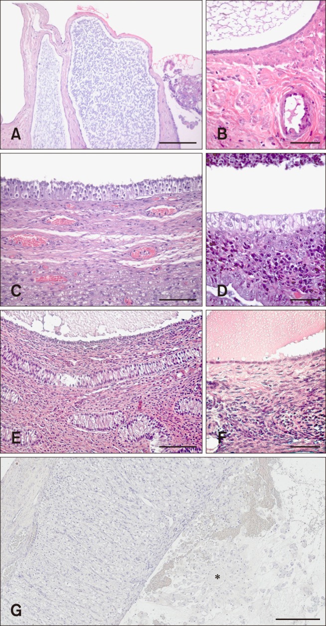
Fig. 1 Histological classification of canine ovarian cysts. (A) Cysts of subsurface epithelial structure (SES): Oval structures located in the ovarian cortex surrounded by smooth muscle. (B) Cyst of SES: Lined by low epithelium without a basement membrane. (C) Follicular cyst (FC): Lined by granulosa cells with a “picket fence” appearance and persistence of recognizable theca interna with blood vessels. (D) FC: Lined by granulosa cells with a “picket fence” appearance and variably sized plaques of luteinized tissue as cystic content. (E) Cystic rete ovarii (CRO): Lined by a single cuboidal epithelium. (F) CRO: Lined by a single cuboidal ciliated epithelium. (G) Lutein cyst (LC): Lined by several layers of granulosa-lutein cells and a thin layer of fibrin on the luminal surface. Capsule of compressed ovarian stroma and vascular connective tissue. Central cavity contains residues of an oocyte (*). H&E stain (A–G). Scale bars = 100 µm (A, C, and E), 50 µm (B, D, and F), 300 µm (G).
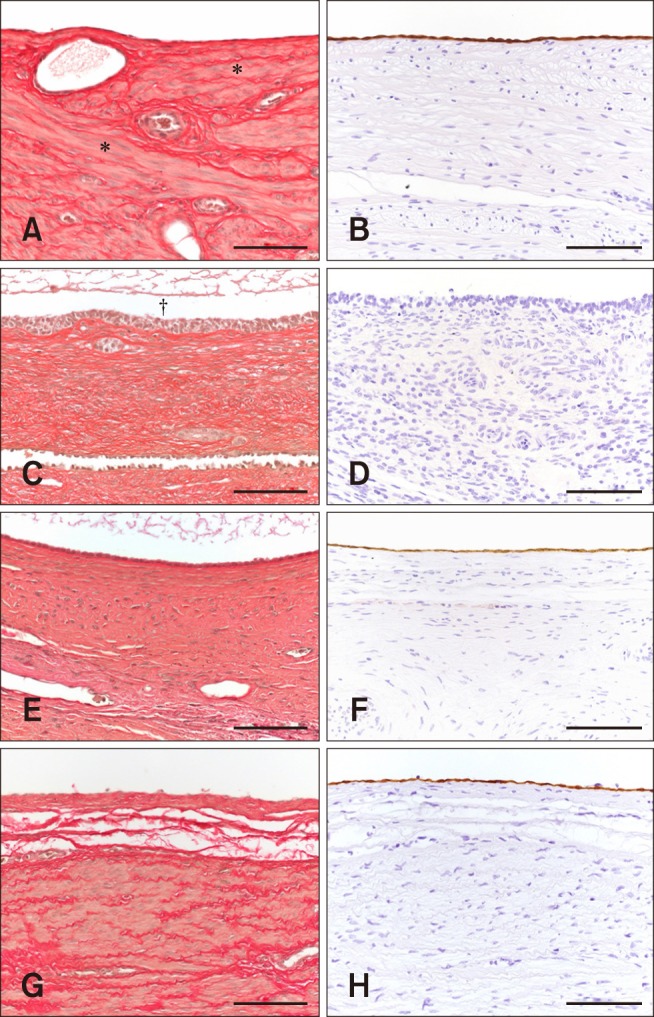
Fig. 2 Histological and immunohistochemical classifications of canine ovarian cysts. (A) Cysts of subsurface epithelial structure (SES): smooth muscle cells surrounding the low epithelium cells that line the cyst (*). (B) Cyst of SES: Low epithelium cells show a strong positive reaction to cytokeratin. (C) Follicular cyst (FC): cavity of the cyst (†). Elastic fibers surrounding the granulosa cells with a picket-fence appearance that lines the cyst. (D) FC: Lined by granulosa cells, which show no reaction to cytokeratin compared to cysts of SES and cystic rete ovarii (CRO). (E) CRO: Lined by cuboidal epithelium cells and surrounded by elastic fibers. (F) CRO: Lining epithelium cells show a weak positive reaction to cytokeratin. (G) Non-classifiable cyst (NCC): Smooth muscle cells and elastic fibers surround the low epithelium cells that line the cyst. (H) NCC: Lining epithelium cells show a positive reaction to cytokeratin. Elastica van Gieson stain (A, C, E, and G). Mouse anti-cytokeratin antibody stain (B, D, F, and H). Scale bars = 100 µm (A–H).
Reference
-
1. Akihara Y, Shimoyama Y, Kawasako K, Komine M, Hirayama K, Kagawa Y, Omachi T, Matsuda K, Okamoto M, Kadosawa T, Taniyama H. Immunohistochemical evaluation of canine ovarian cysts. J Vet Med Sci. 2007; 69:1033–1037. PMID: 17984590.
Article2. Amweg AN, Rodríguez FM, Huber E, Marelli BE, Salvetti NR, Rey F, Ortega HH. Role of glucocorticoids in cystic ovarian disease: expression of glucocorticoid receptor in the bovine ovary. Cells Tissues Organs. 2016; 201:138–147. PMID: 26677854.
Article3. Andersen AC, Simpson ME. Pathology of the ovary and genital tract. In : Andersen AC, Simpson ME, editors. The Ovary and Reproductive Cycle of the Dog (Beagle). Los Altos: Geron-X;1973. p. 246–272.4. Arlt SP, Haimerl P. Cystic ovaries and ovarian neoplasia in the female dog - a systematic review. Reprod Domest Anim. 2016; 51(Suppl 1):3–11.
Article5. Arlt SP, Spankowsky S, Heuwieser W. Follicular cysts and prolonged oestrus in a female dog after administration of a deslorelin implant. N Z Vet J. 2011; 59:87–91. PMID: 21409735.
Article6. Bostedt H, Jung C, Wehrend A, Boryzcko Z. [Clinical and endocrinological findings of bitches with ovarian cyst syndrome]. Schweiz Arch Tierheilkd. 2013; 155:543–550. German. PMID: 24091229.7. Bostedt H, Tammer I, Hecker BR. Infertility in the breeding bitch: a short review. Tierärztl Prax. 1999; 27:179–185.8. Bowen RA, Olson PN, Young S, Withrow SJ. Efficacy and toxicity of tamoxifen citrate for prevention and termination of pregnancy in bitches. Am J Vet Res. 1988; 49:27–31. PMID: 3354962.9. Chuffa LG, Lupi Júnior LA, da Maia Lima AF. Sex steroid receptors and apoptosis-related proteins are differentially expressed in polycystic ovaries of adult dogs. Tissue Cell. 2016; 48:10–17. PMID: 26767421.
Article10. Dow C. Ovarian abnormalities in the bitch. J Comp Pathol. 1960; 70:59–69. PMID: 13817873.
Article11. Ervin E, Homans P. Giant ovarian cyst. Compend Contin Educ Pract Vet. 1986; 8:698–700.12. Fontbonne A. Infertility in the bitch. In : Proceedings of the 31th World Small Animal Veterinary Association (WSAVA) Congress; 11 October 2006; 11 October 2006.13. Groeger S, Weiss R, Trasch K, Wehrend A. Significance of the bacteriological results from vaginal swabs taken from bitches with overt pyometra. Kleintierpraxis. 2007; 52:426–428.14. Gulliver LS, Hurst PR. Repeat estradiol exposure differentially regulates protein expression patterns for estrogen receptor and E-cadherin in older mouse ovarian surface epithelium: implications for replacement and adjuvant hormone therapies? Steroids. 2012; 77:674–685. PMID: 22406420.
Article15. Johnston SD, Kustritz MV, Olson PS. Disorders of the canine ovary. Canine and Feline Theriogenology. Philadelphia: Saunders;2001. p. 193–205.16. Kennedy PC, Cullen JM, Edwards JF, Goldschmidt MH, Larsen S, Munson L, Nielsen S. Cysts in and around the ovary. In : Kennedy PC, editor. Histological Classification of Tumors of the Genital System of Domestic Animals, Vol. IV. 2nd ed. Washington: Armed Forces Institute of Pathology;1998. p. 29–31.17. Kesler DJ, Garverick HA. Ovarian cysts in dairy cattle: a review. J Anim Sci. 1982; 55:1147–1159. PMID: 6757234.
Article18. Knauf Y, Bostedt H, Failing K, Knauf S, Wehrend A. Gross pathology and endocrinology of ovarian cysts in bitches. Reprod Domest Anim. 2014; 49:463–468. PMID: 24698026.
Article19. Knauf Y, Failing K, Knauf S, Wehrend A. [Treatment of bitches with ovarian cysts using human chorionic gonadotropin-releasing hormone analogue. A case series of 30 bitches]. Tierarztl Prax Ausg K Kleintiere Heimtiere. 2013; 41:93–100. German. PMID: 23608964.20. Knauf Y, Wehrend A. [Ovarian cysts in the bitch]. Tierarztl Prax Ausg K Kleintiere Heimtiere. 2010; 38:333–340. German. PMID: 22215319.21. Lecke SB, Mattei F, Morsch DM, Spritzer PM. Abdominal subcutaneous fat gene expression and circulating levels of leptin and adiponectin in polycystic ovary syndrome. Fertil Steril. 2011; 95:2044–2049. PMID: 21419406.
Article22. Marino G, Mannarino C, Di Prima ML, Rizzo S, Zanghi A. Stromal cysts in the canine ovary. In : Proceedings of the 27th Meeting of the European Society of Veterinary Pathology and European College of Veterinary Pathologists; 9-12 September 2009; Olsztyn-Kraków, Poland..23. McEntee K. Cysts in and around the ovary. Reproductive Pathology of Domestic Mammals. San Diego: Academic Press;1990. p. 52–67.24. Miller DM, McCrory VS, Anderson WI. Polycystic ovarian tissue in a spayed bitch. Mod Vet Pract. 1983; 64:749.25. Olson PN, Wrigley RH, Husted PW, Bowen RA, Nett TA. Persistent estrus in the bitch. In : Ettinger SJ, editor. Textbook of Veterinary Internal Medicine, Vol. 2. Philadelphia: WB Saunders;1989. p. 1792–1796.26. Ortega-Pacheco A, Segura-Correa JC, Jimenez-Coello M, Linde Forsberg C. Reproductive patterns and reproductive pathologies of stray bitches in the tropics. Theriogenology. 2007; 67:382–390. PMID: 17007916.
Article27. Radavelli-Bagatini S, Blair AR, Proietto J, Spritzer PM, Andrikopoulos S. The New Zealand obese mouse model of obesity insulin resistance and poor breeding performance: evaluation of ovarian structure and function. J Endocrinol. 2011; 209:307–315. PMID: 21429962.
Article28. Sant'Ana F, Reis Junior JL, Blume GR, Gimeno EJ, Rey F, Ortega HH. Immunohistochemical expression of growth factors in the follicular wall of normal and cystic ovaries of sows. Reprod Domest Anim. 2015; 50:327–332. PMID: 25676567.29. Schlafer DH, Miller RB. Pathology of the ovary (nondevelopmental lesions). In : Maxie MG, Jubb KVF, Kennedy PC, Palmer N, editors. Jubb, Kennedy, and Palmer's Pathology of Domestic Animals, Vol. 3. Philadelphia: Elsevier Saunders;2007. p. 431–563.30. Schwarz H, Geyer S, Rüsse M, Hänichen T. [Intoxication caused by estrogen administration in the bitch]. Tierarztl Prax. 1982; 10:393–402. German. PMID: 7179267.31. Shille VM, Calderwood-Mays MB, Thatcher MJ. Infertility in a bitch associated with short interestrus intervals and cystic follicles: a case report. J Am Anim Hosp Assoc. 1984; 20:171–176.32. Songsasen N, Fickes A, Pukazhenthi BS, Wildt DE. Follicular morphology, oocyte diameter and localisation of fibroblast growth factors in the domestic dog ovary. Reprod Domest Anim. 2009; 44(Suppl 2):65–70. PMID: 19754538.
Article33. Suttorp M, Hoffmann B, Sippell WG. Prevention of oestradiol-associated toxicosis in a dalmatian by early intervention with granulocyte colony-stimulating factor. Vet Rec. 2002; 151:244–245. PMID: 12219904.
Article34. Tammer I, Blendinger K, Sobiraj A, Bostedt H. [The use of exfoliative vaginal cytology for the gynecological evaluation of the bitch]. Tierarztl Prax. 1994; 22:199–207. German. PMID: 7519369.35. Trasch K, Wehrend A, Bostedt H. Follow-up examinations of bitches after conservative treatment of pyometra with the antigestagen aglepristone. J Vet Med A Physiol Pathol Clin Med. 2003; 50:375–379. PMID: 14633233.
Article36. Volk KM, Pogrebna VV, Roberts JA, Zachry JE, Blythe SN, Toporikova N. High-fat, high-sugar diet disrupts the preovulatory hormone surge and induces cystic ovaries in cycling female rats. J Endocr Soc. 2017; 1:1488–1505. PMID: 29308444.
Article37. Weiss E. Weibliche Geschlechtsorgane. In : Dahme E, Hafner-Marx A, editors. [Grundriss der Speziellen Pathologischen Anatomie der Haustiere, Vol. 6]. Stuttgart: Enke;2007. p. 213–232. German.
- Full Text Links
-
- Actions
-
Cited
- CITED
-
- Close
- Share
-
- Similar articles
-
- Natural Course and Treatment of Fetal Ovarian Cysts
- A Case of Adenocarcinoma Arising from Dermoid Cyst of the Ovary
- A Case of Antenatally Diagnosed Fetal Ovarian Cyst
- Two Cases of Ovarian Dermoid Cyst Extended into the Urinary Bladder
- Transvaginal sonography guided aspiration of ovarian cyst suspected torsion in early pregnancy: A case report
