Korean J Orthod.
2016 Nov;46(6):345-355. 10.4041/kjod.2016.46.6.345.
Relationship between maturation indices and morphology of the midpalatal suture obtained using cone-beam computed tomography images
- Affiliations
-
- 1Department of Orthodontics, School of Dentistry, Wonkwang University, Iksan, Korea. jjhdent@wonkwang.ac.kr
- 2Wonkwang Dental Research Institute, School of Dentistry, Wonkwang University, Iksan, Korea.
- 3Department of Orthodontics, Wonkwang University Daejeon Dental Hospital, Daejeon, Korea.
- KMID: 2426678
- DOI: http://doi.org/10.4041/kjod.2016.46.6.345
Abstract
OBJECTIVE
The purpose of this study was to determine whether predicting maturation of the midpalatal suture is possible by classifying its morphology on cone-beam computed tomography (CBCT) images and to investigate relationships with other developmental age indices.
METHODS
The morphology of the midpalatal suture was assessed by using CBCT images of 99 patients. Axial plane images of the midpalatal suture were classified into five stages according to the classification scheme. To make the assessment more accurate, the morphology and fusion of the midpalatal suture were additionally investigated on coronal cross-sectional planar images and volume-rendered images. Bone age was evaluated using the hand and wrist method (HWM) and cervical vertebrae method (CVM); dental age (Hellman's index), sex, and chronological age were also assessed. To evaluate relationships among variables, Spearman's rho rank test was performed along with crosstabs using contingency coefficients.
RESULTS
The HWM and CVM showed strong correlations with the maturation stage of the midpalatal suture, while other indices showed relatively weak correlations (p < 0.01). Through crosstabs, the HWM and CVM showed high association values with CBCT stage; the HWM demonstrated slightly higher values (p < 0.0001). Based on the HWM, the midpalatal suture was not fused until stage 6 in both sexes.
CONCLUSIONS
Among developmental age indices, the HWM and CVM showed strong correlations and high associations, suggesting that they can be useful in assessing maturation of the midpalatal suture.
MeSH Terms
Figure
-

Figure 1 Head reorientation and setting of the axial cross-sectional planar view. A, An axial plane view; B, a coronal plane view; C, a sagittal plane view; and D, a midsagittal plane view.
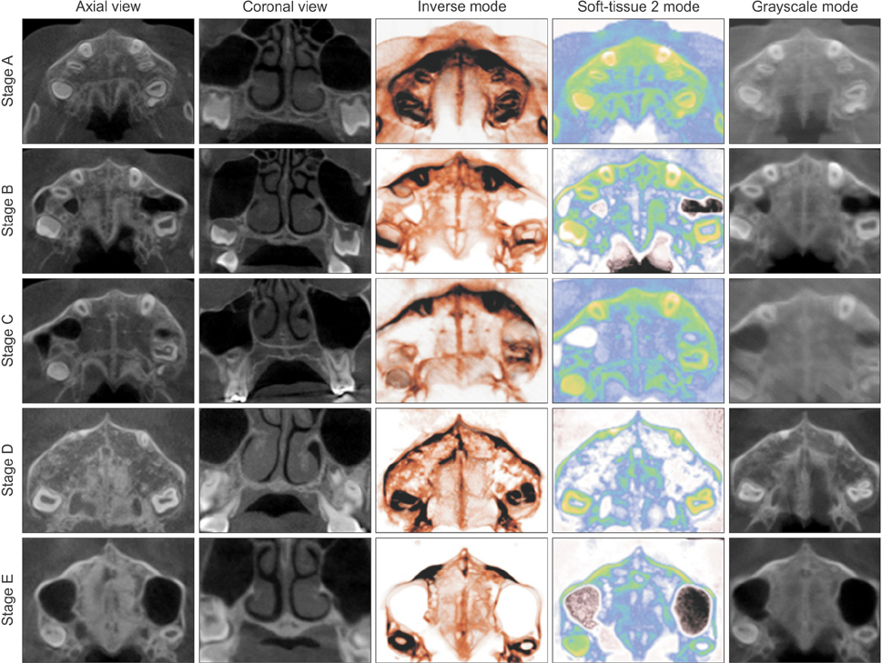
Figure 2 Cone-beam computed tomography images of the midpalatal suture and volume-rendered images according to maturation stage.
Cited by 1 articles
-
Stability of dental, alveolar, and skeletal changes after miniscrew-assisted rapid palatal expansion
Hyun-Mook Lim, Young-Chel Park, Kee-Joon Lee, Kyung-Ho Kim, Yoon Jeong Choi
Korean J Orthod. 2017;47(5):313-322. doi: 10.4041/kjod.2017.47.5.313.
Reference
-
1. Gill D, Naini F, McNally M, Jones A. The management of transverse maxillary deficiency. Dent Update. 2004; 31:516–518. 521–523.
Article2. Melsen B. Palatal growth studied on human autopsy material. A histologic microradiographic study. Am J Orthod. 1975; 68:42–54.3. Suri L, Taneja P. Surgically assisted rapid palatal expansion: a literature review. Am J Orthod Dentofacial Orthop. 2008; 133:290–302.
Article4. Wehrbein H, Merz BR, Diedrich P. Palatal bone support for orthodontic implant anchorage--a clinical and radiological study. Eur J Orthod. 1999; 21:65–70.
Article5. Lee KJ, Park YC, Park JY, Hwang WS. Miniscrew-assisted nonsurgical palatal expansion before orthognathic surgery for a patient with severe mandibular prognathism. Am J Orthod Dentofacial Orthop. 2010; 137:830–839.
Article6. Fishman LS. Radiographic evaluation of skeletal maturation. A clinically oriented method based on hand-wrist films. Angle Orthod. 1982; 52:88–112.7. Fishman LS. Maturational patterns and prediction during adolescence. Angle Orthod. 1987; 57:178–193.8. Hassel B, Farman AG. Skeletal maturation evaluation using cervical vertebrae. Am J Orthod Dentofacial Orthop. 1995; 107:58–66.
Article9. Hellman M. The process of dentition and its effects on occlusion. Dent Cosmos. 1923; 65:1329–1344.10. Angelieri F, Cevidanes LH, Franchi L, Gonçalves JR, Benavides E, McNamara JA Jr. Midpalatal suture maturation: classification method for individual assessment before rapid maxillary expansion. Am J Orthod Dentofacial Orthop. 2013; 144:759–769.
Article11. Timms DJ, Vero D. The relationship of rapid maxillary expansion to surgery with special reference to midpalatal synostosis. Br J Oral Surg. 1981; 19:180–196.
Article12. Epker BN, Wolford LM. Transverse maxillary deficiency dentofacial deformities: integrated orthodontic and surgical correction. St. Louis: Mosby;1980.13. Mossaz CF, Byloff FK, Richter M. Unilateral and bilateral corticotomies for correction of maxillary transverse discrepancies. Eur J Orthod. 1992; 14:110–116.
Article14. Mommaerts MY. Transpalatal distraction as a method of maxillary expansion. Br J Oral Maxillofac Surg. 1999; 37:268–272.
Article15. Alpern MC, Yurosko JJ. Rapid palatal expansion in adults with and without surgery. Angle Orthod. 1987; 57:245–263.16. Fishman LS. Chronological versus skeletal age, an evaluation of craniofacial growth. Angle Orthod. 1979; 49:181–189.17. Flores-Mir C, Nebbe B, Major PW. Use of skeletal maturation based on hand-wrist radiographic analysis as a predictor of facial growth: a systematic review. Angle Orthod. 2004; 74:118–124.18. Kwak KH, Kim SS, Kim YI, Kim YD. Quantitative evaluation of midpalatal suture maturation via fractal analysis. Korean J Orthod. 2016; 46:323–330.
Article19. Proffit WR, Fields HW, Sarver DM. Contemporary orthodontics. 5th ed. St. Louis: Elsevier/Mosby;2013.20. Johnston FE, Hufham HP Jr, Moreschi AF, Terry GD. Skeletal maturation and cephalofacial development. Angle Orthod. 1965; 35:1–11.21. Danaei SM, Karamifar A, Sardarian A, Shahidi S, Karamifar H, Alipour A, et al. Measuring agreement between cervical vertebrae and hand-wrist maturation in determining skeletal age: Reassessing the theory in patients with short stature. Am J Orthod Dentofacial Orthop. 2014; 146:294–298.
Article22. Wong RW, Alkhal HA, Rabie AB. Use of cervical vertebral maturation to determine skeletal age. Am J Orthod Dentofacial Orthop. 2009; 136:484.e1–484.e6.
Article23. García-Fernandez P, Torre H, Flores L, Rea J. The cervical vertebrae as maturational indicators. J Clin Orthod. 1998; 32:221–225.24. Beit P, Peltomäki T, Schätzle M, Signorelli L, Patcas R. Evaluating the agreement of skeletal age assessment based on hand-wrist and cervical vertebrae radiography. Am J Orthod Dentofacial Orthop. 2013; 144:838–847.
Article25. Mellion ZJ, Behrents RG, Johnston LE Jr. The pattern of facial skeletal growth and its relationship to various common indexes of maturation. Am J Orthod Dentofacial Orthop. 2013; 143:845–854.
Article26. Koudstaal MJ, Poort LJ, van der Wal KG, Wolvius EB, Prahl-Andersen B, Schulten AJ. Surgically assisted rapid maxillary expansion (SARME): a review of the literature. Int J Oral Maxillofac Surg. 2005; 34:709–714.
Article27. Magnusson A, Bjerklin K, Nilsson P, Jönsson F, Marcusson A. Nasal cavity size, airway resistance, and subjective sensation after surgically assisted rapid maxillary expansion: a prospective longitudinal study. Am J Orthod Dentofacial Orthop. 2011; 140:641–651.
Article28. Ghoneima A, Abdel-Fattah E, Hartsfield J, El-Bedwehi A, Kamel A, Kula K. Effects of rapid maxillary expansion on the cranial and circum-maxillary sutures. Am J Orthod Dentofacial Orthop. 2011; 140:510–519.
Article29. N'Guyen T, Gorse FC, Vacher C. Anatomical modifications of the mid palatal suture during ageing: a radiographic study. Surg Radiol Anat. 2007; 29:253–259.
- Full Text Links
-
- Actions
-
Cited
- CITED
-
- Close
- Share
-
- Similar articles
-
- Evaluation of the Midpalatal Suture Maturation in Young Koreans Using Cone-Beam Computed Tomography
- Evaluation of Midpalatal Suture Maturation using Cone-Beam Computed Tomography in Children and Adolescents
- Assessment of Midpalatal Suture Maturation by Skeletal Maturity on Hand Wrist Radiographs
- Quantitative evaluation of midpalatal suture maturation via fractal analysis
- Utility of the computed tomography indices on cone beam computed tomography images in the diagnosis of osteoporosis in women
