Clin Orthop Surg.
2018 Dec;10(4):500-507. 10.4055/cios.2018.10.4.500.
Reliability of the EOS Imaging System for Assessment of the Spinal and Pelvic Alignment in the Sagittal Plane
- Affiliations
-
- 1Department of Orthopedic Surgery, Konyang University Hospital, Daejeon, Korea. oeo-oeoeo@hanmail.net
- 2Department of Radiology, Konyang University Hospital, Daejeon, Korea.
- 3Department of Preventive Medicine, Konyang University College of Medicine, Daejeon, Korea.
- KMID: 2426537
- DOI: http://doi.org/10.4055/cios.2018.10.4.500
Abstract
- BACKGROUND
The sagittal alignment of the spine and pelvis is not only closely related to the overall posture of the body but also to the evaluation and treatment of spine disease. In the last few years, the EOS imaging system, a new low-dose radiation X-ray device, became available for sagittal alignment assessment. However, there has been little research on the reliability of EOS. The purpose of this study was to evaluate the intrarater and interrater reliability of EOS for the sagittal alignment assessment of the spine and pelvis.
METHODS
Records of 46 patients were selected from the EOS recording system between November 2016 and April 2017. The exclusion criteria were congenital spinal anomaly and deformity, and previous history of spine and pelvis operation. Sagittal parameters of the spine and pelvis were measured by three examiners three times each using both manual and EOS methods. Means comparison t-test, Pearson bivariate correlation analysis, and reliability analysis by intraclass correlation coefficients (ICCs) for intrarater and interrater reliability were performed using R package "irr."
RESULTS
We found excellent intrarater and interrater reliability of EOS measurements. For intrarater reliability, the ICC ranged from 0.898 to 0.982. For interrater reliability, the ICC ranged from 0.794 to 0.837. We used a paired t-test to compare the values measured by manual and EOS methods: there was no statistically significant difference between the two methods. Correlation analysis also showed a statistically significant positive correlation.
CONCLUSIONS
EOS showed excellent reliability for assessment of the sagittal alignment of the spine and pelvis.
MeSH Terms
Figure
-
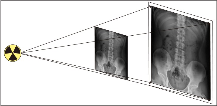
Fig. 1 Distortion caused by conical projection from the center to the edges of radiograph, which increases the scale of error for structures farther from the central region.
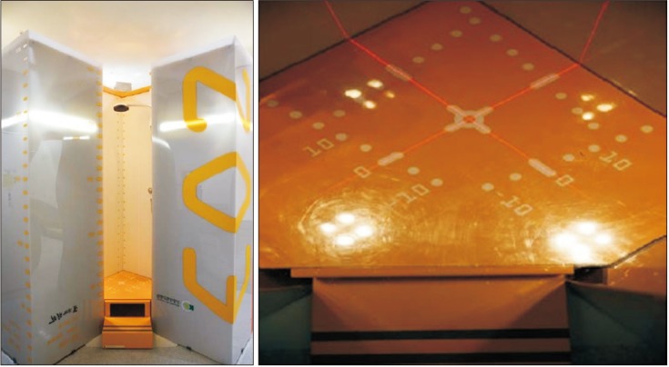
Fig. 2 EOS system cabin to scan the patient in a standing position in two orthogonal planes.
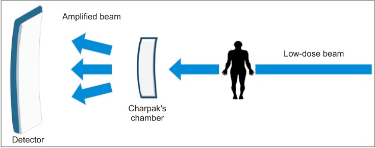
Fig. 3 In EOS, when a low-dose X-ray beam passes through the subject and Charpak's chamber, the flow of photons increases in the chamber, amplifying the low-dose X-rays.
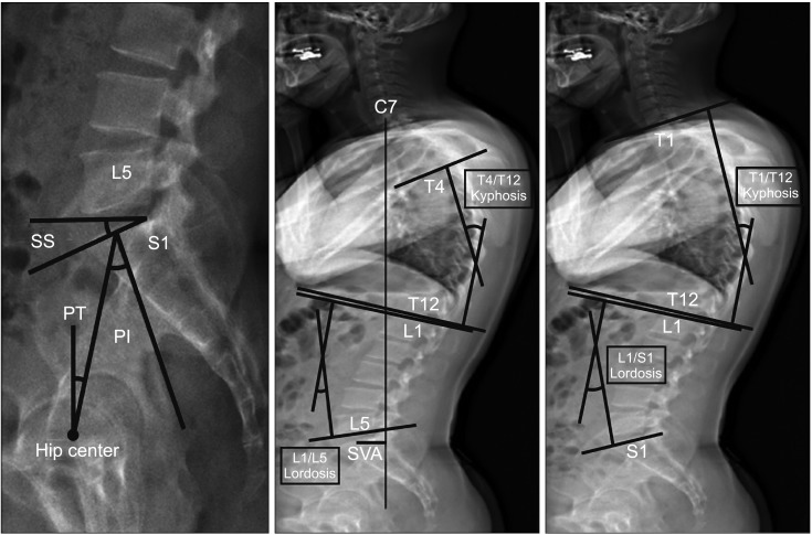
Fig. 4 The following eight items were measured using the EOS imaging system: pelvic incidence (PI), sacral slope (SS), sagittal pelvic tilt (PT), sagittal vertical axis (SVA), T1/T12 and T4/T12 kyphosis, and L1/S1 and L1/L5 lordosis.
Reference
-
1. Lee CS, Chung SS, Kang KC, Park SJ, Shin SK. Normal patterns of sagittal alignment of the spine in young adults radiological analysis in a Korean population. Spine (Phila Pa 1976). 2011; 36(25):E1648–E1654. PMID: 21394071.
Article2. Korovessis PG, Stamatakis MV, Baikousis AG. Reciprocal angulation of vertebral bodies in the sagittal plane in an asymptomatic Greek population. Spine (Phila Pa 1976). 1998; 23(6):700–704. PMID: 9549792.
Article3. Van Royen BJ, Toussaint HM, Kingma I, et al. Accuracy of the sagittal vertical axis in a standing lateral radiograph as a measurement of balance in spinal deformities. Eur Spine J. 1998; 7(5):408–412. PMID: 9840475.
Article4. Vedantam R, Lenke LG, Keeney JA, Bridwell KH. Comparison of standing sagittal spinal alignment in asymptomatic adolescents and adults. Spine (Phila Pa 1976). 1998; 23(2):211–215. PMID: 9474728.
Article5. Somoskeoy S, Tunyogi-Csapo M, Bogyo C, Illes T. Accuracy and reliability of coronal and sagittal spinal curvature data based on patient-specific three-dimensional models created by the EOS 2D/3D imaging system. Spine J. 2012; 12(11):1052–1059. PMID: 23102842.6. Kuklo TR, Potter BK, Polly DW Jr, O'Brien MF, Schroeder TM, Lenke LG. Reliability analysis for manual adolescent idiopathic scoliosis measurements. Spine (Phila Pa 1976). 2005; 30(4):444–454. PMID: 15706343.
Article7. Deschenes S, Charron G, Beaudoin G, et al. Diagnostic imaging of spinal deformities: reducing patients radiation dose with a new slot-scanning X-ray imager. Spine (Phila Pa 1976). 2010; 35(9):989–994. PMID: 20228703.8. Nobel Media. The Nobel Prize in Physics 1992 [Internet]. London: Nobel Media;1992. cited 2018 Oct 5. Available from: https://www.nobelprize.org/prizes/uncategorized/thenobel-prize-in-physics-1992-1992.9. Dubousset J, Charpak G, Dorion I, et al. A new 2D and 3D imaging approach to musculoskeletal physiology and pathology with low-dose radiation and the standing position: the EOS system. Bull Acad Natl Med. 2005; 189(2):287–297. PMID: 16114859.10. Escott BG, Ravi B, Weathermon AC, et al. EOS low-dose radiography: a reliable and accurate upright assessment of lower-limb lengths. J Bone Joint Surg Am. 2013; 95(23):e1831–e1837. PMID: 24306706.11. Wybier M, Bossard P. Musculoskeletal imaging in progress: the EOS imaging system. Joint Bone Spine. 2013; 80(3):238–243. PMID: 23177915.
Article12. Lee KM, Chung CY, Park MS, Lee SH, Cho JH, Choi IH. Reliability and validity of radiographic measurements in hindfoot varus and valgus. J Bone Joint Surg Am. 2010; 92(13):2319–2327. PMID: 20926727.
Article13. McGraw KO, Wong SP. Forming inferences about some intraclass correlation coefficients. Psychol Methods. 1996; 1(1):30–46.
Article14. Bonett DG. Sample size requirements for estimating intraclass correlations with desired precision. Stat Med. 2002; 21(9):1331–1335. PMID: 12111881.
Article15. Fleiss JL. Statistical methods for rates and proportions. 2nd ed. New York, NY: Wiley;1981.16. Dubousset J, Charpak G, Skalli W, de Guise J, Kalifa G, Wicart P. Skeletal and spinal imaging with EOS system. Arch Pediatr. 2008; 15(5):665–666. PMID: 18582707.
- Full Text Links
-
- Actions
-
Cited
- CITED
-
- Close
- Share
-
- Similar articles
-
- Introduction of a New Skeletal Imaging Instrument: The Low Radiating-Dose EOS System
- Sagittal Parameters of Spine and Pelvis in Young Adults Using the EOS Imaging System: Prospective Study of 92 Asymptomatic Subjects
- Comparison of Whole Spine Sagittal Alignment in Patients with Spinal Disease between EOS Imaging System versus Conventional Whole Spine X-ray
- Radiologic Findings of Pelvic Parameters Related to Sagittal Balance
- Spinal Deformity Surgery: A Critical Review of Alignment and Balance
