Clin Endosc.
2013 Jul;46(4):407-409.
A Case of Squamous Metaplasia of the Stomach
- Affiliations
-
- 1Department of Internal Medicine, Pusan National University School of Medicine, Busan, Korea. doc0224@pusan.ac.kr
- 2Department of Pathology, Pusan National University School of Medicine, Busan, Korea.
- 3Department of Internal Medicine, Good Samsun Hospital, Busan, Korea.
Abstract
- Intestinal metaplasia of the stomach is a common metaplastic lesion associated with chronic gastritis and mucosal atrophy. However, squamous metaplasia is a comparatively rare condition. On endoscopy, squamous metaplasia is usually observed as a whitish mucosal lesion in the lesser curvature of the cardiac region of the stomach. When Lugol's iodine solution is applied, the lesion stains brown in the same way as normal esophageal mucosa. We report a case of 79-year-old man with a whitish flat lesion in the lesser curvature of the cardiac region on surveillance endoscopy after endoscopic treatment of gastric adenoma. The endoscopic biopsy showed stratified squamous epithelial mucosa.
MeSH Terms
Figure
-
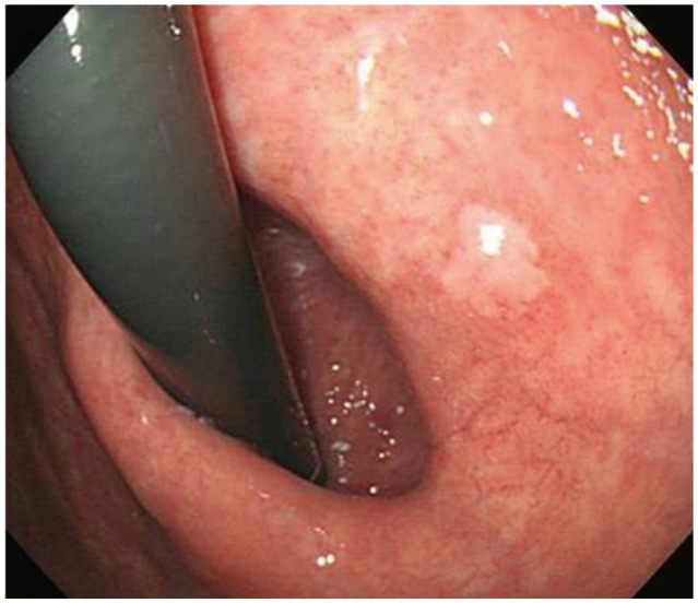
Fig. 1 A surveillance endoscopy after endoscopic treatment of gastric adenoma. An area of whitish lesion is found in the lesser curvature of the cardiac region of the stomach.
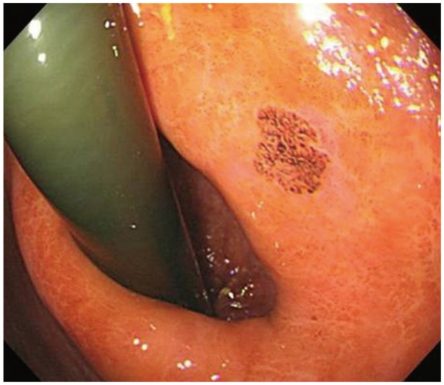
Fig. 2 Chromoendoscopy using Lugol's iodine solution. The whitish lesion is stained brown with Lugol's iodine solution.
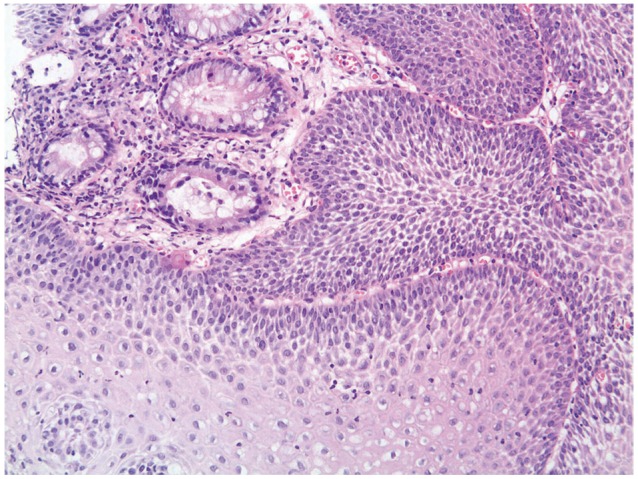
Fig. 3 Histopathologic finding. The biopsy from the white area revealed stratified squamous epithelial mucosa (H&E stain, ×100).
Reference
-
1. Vaughan WP, Straus FH 2nd, Paloyan D. Squamous carcinoma of the stomach after luetic linitis plastica. Gastroenterology. 1977; 72(5 Pt 1):945–948. PMID: 191328.
Article2. Watson GW, Flint ER, Stewart MJ. Hyperplastic tuberculosis of the stomach causing hour-glass deformity, with complete squamous metaplasia of the upper loculus. Br J Surg. 1936; 24:333–340.
Article3. Yamagiwa H, Yosimura H, Nishii M, Moriyama S. Squamous metaplasia of the gastric mucosa associated with an aberrant pancreas. Gan No Rinsho. 1987; 33:1929–1932. PMID: 3430744.4. Takeda H, Nagashima R, Goto T, Shibata Y, Shinzawa H, Takahashi T. Endoscopic observation of squamous metaplasia of the stomach: a report of two cases. Endoscopy. 2000; 32:651–653. PMID: 10935797.
Article5. Boswell JT, Helwig EB. Squamous cell carcinoma and adenoacanthoma of the stomach. A clinicopathologic study. Cancer. 1965; 18:181–192. PMID: 14254074.6. Fennerty MB, Sampliner RE, McGee DL, Hixson LJ, Garewal HS. Intestinal metaplasia of the stomach: identification by a selective mucosal staining technique. Gastrointest Endosc. 1992; 38:696–698. PMID: 1282115.
Article7. Rerknimitr R. Practical use of digital chromoendoscopy for GI tract diseases including GERD. Thai J Gastroenterol. 2009; 9:147–159.8. Parks RE. Squamous neoplasms of the stomach. Am J Roentgenol Radium Ther Nucl Med. 1967; 101:447–449.
Article9. Wood DA. Adenoacanthoma of the pyloric end of the stomach. Arch Pathol. 1943; 36:177–189.
