J Clin Neurol.
2012 Jun;8(2):123-129.
Effect of Cardiac Function on Cognition and Brain Structural Changes in Dementia
- Affiliations
-
- 1Department of Neurology, Chungnam National University College of Medicine, Daejeon, Korea. aelee@cnu.ac.kr
- 2Department of Cardiology, Chungnam National University College of Medicine, Daejeon, Korea.
Abstract
- BACKGROUND AND PURPOSE
Cardiovascular risk factors are considered to also be risk factors for dementia. Recent studies have shown that the prevalence of cognitive dysfunction is high in patients with cardiac diseases. However, few studies have investigated the influence of cardiac function on cognition and brain structural changes in dementia. The aims of this study were to determine the relationship between cardiac and cognitive function, and to characterize any structural changes in the brain that could be caused by cardiac function in patients with dementia.
METHODS
Dementia patients (n=93) were recruited prospectively with checking for the presence of vascular risk factors such as hypertension. Cognitive function was measured by the Mini-Mental State Examination, modified Mini-Mental State test, and Korean version of the Dementia Rating Scale. Brain magnetic resonance imaging was conducted to evaluate the cerebral white-matter changes (WMC), ventricular dilation, and cortical and hippocampal atrophy. Cardiac function was evaluated using two-dimensional echocardiography. We divided the patients into two groups according to the presence (+) or absence (-) of WMC.
RESULTS
In the entire cohort, the size of the left atrium (LA) was positively correlated with the degree of WMC, irrespective of age (p<0.05). The LA was larger in the WMC (+) group (n=42) than in the WMC (-) group. General cognitive function was significantly lower in the WMC (+) group than in the WMC (-) group. Subjects with an enlarged LA tended to exhibit lower cognitive function and more-severe cerebral WMC.
CONCLUSIONS
Cardiac dysfunction represented by LA enlargement could be related to cognitive decline and WMC of the brain resulting from impairment of the cerebral hemodynamic process in dementia.
MeSH Terms
Figure
-
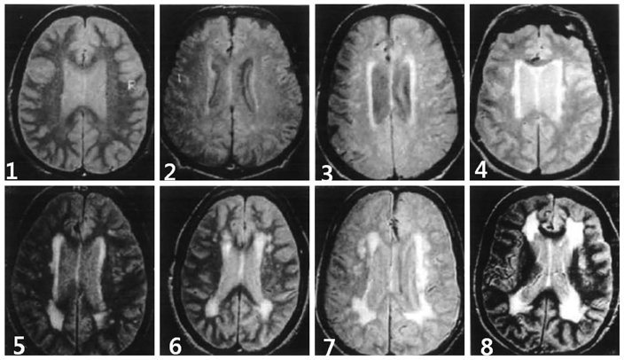
Fig. 1 Severity of the cerebral white matter changes, as determined using the Cardiovascular Health Study Scale (score 1-8).

Fig. 2 Degree of cortical atrophy, as determined using the Cardiovascular Health Study Scale (score 1-8).

Fig. 3 Degree of ventricular enlargement, as determined using the Cardiovascular Health Study Scale (score 1-8).

Fig. 4 Severity of medial temporal lobe atrophy, as determined using Scheltens' Scale (score 0-4).

Fig. 5 Comparison of cardiac and cognitive functions between WMC-positive (+) and WMC-negative (-) groups. CO: cardiac output, EF: ejection fraction, LA: left atrium, MMSE: the Mini-Mental State Examination, WMC: white matter changes, 3MS: the modified Mini-Mental State test.
Reference
-
1. Statistics Korea. 2010. Available from: URL: http://www.kostat.go.kr.2. McCullagh CD, Craig D, McIlroy SP, Passmore AP. Risk factors for dementia. Adv Psychiatr Treat. 2001. 7:24–31.
Article3. Fillit H, Nash DT, Rundek T, Zuckerman A. Cardiovascular risk factors and dementia. Am J Geriatr Pharmacother. 2008. 6:100–118.
Article4. Zuccalà G, Cattel C, Manes-Gravina E, Di Niro MG, Cocchi A, Bernabei R. Left ventricular dysfunction: a clue to cognitive impairment in older patients with heart failure. J Neurol Neurosurg Psychiatry. 1997. 63:509–512.
Article5. Vogels RL, van der Flier WM, van Harten B, Gouw AA, Scheltens P, Schroeder-Tanka JM, et al. Brain magnetic resonance imaging abnormalities in patients with heart failure. Eur J Heart Fail. 2007. 9:1003–1009.
Article6. Suwa M, Ito T. Correlation between cognitive impairment and left ventricular diastolic dysfunction in patients with cardiovascular diseases. Int J Cardiol. 2009. 136:351–354.
Article7. McKhann G, Drachman D, Folstein M, Katzman R, Price D, Stadlan EM. Clinical diagnosis of Alzheimer's disease: report of the NINCDS-ADRDA Work Group under the auspices of Department of Health and Human Services Task Force on Alzheimer's Disease. Neurology. 1984. 34:939–944.
Article8. Erkinjuntti T, Inzitari D, Pantoni L, Wallin A, Scheltens P, Rockwood K, et al. Research criteria for subcortical vascular dementia in clinical trials. J Neural Transm Suppl. 2000. 59:23–30.
Article9. Emre M, Aarsland D, Brown R, Burn DJ, Duyckaerts C, Mizuno Y, et al. Clinical diagnostic criteria for dementia associated with Parkinson's disease. Mov Disord. 2007. 22:1689–1707.
Article10. Teng EL, Chui HC. The Modified Mini-Mental State (3MS) examination. J Clin Psychiatry. 1987. 48:314–318.11. Sohn EH, Lee AY, Park HJ. The validity and reliability of the Korean Modified Mini-Mental State (K-3MS) examination. J Korean Neurol Assoc. 2003. 21:346–356.12. Folstein MF, Folstein SE, McHugh PR. "Mini-mental state". A practical method for grading the cognitive state of patients for the clinician. J Psychiatr Res. 1975. 12:189–198.13. Chey J, Na DR, Park SH, Park EH. The validity and reliability of the Korean dementia rating scale. Korean J Clin Psychol. 1998. 17:247–258.14. Manolio TA, Kronmal RA, Burke GL, Poirier V, O'Leary DH, Gardin JM, et al. Magnetic resonance abnormalities and cardiovascular disease in older adults. The Cardiovascular Health Study. Stroke. 1994. 25:318–327.
Article15. Korf ES, Wahlund LO, Visser PJ, Scheltens P. Medial temporal lobe atrophy on MRI predicts dementia in patients with mild cognitive impairment. Neurology. 2004. 63:94–100.
Article16. Edhouse J, Thakur RK, Khalil JM. ABC of clinical electrocardiography. Conditions affecting the left side of the heart. BMJ. 2002. 324:1264–1267.
Article17. Lang RM, Bierig M, Devereux RB, Flachskampf FA, Foster E, Pellikka PA, et al. Recommendations for chamber quantification. Eur J Echocardiogr. 2006. 7:79–108.
Article18. Bursi F, Rocca WA, Killian JM, Weston SA, Knopman DS, Jacobsen SJ, et al. Heart disease and dementia: a population-based study. Am J Epidemiol. 2006. 163:135–141.
Article19. Kilander L, Andrén B, Nyman H, Lind L, Boberg M, Lithell H. Atrial fibrillation is an independent determinant of low cognitive function: a cross-sectional study in elderly men. Stroke. 1998. 29:1816–1820.
Article20. Abhayaratna WP, Seward JB, Appleton CP, Douglas PS, Oh JK, Tajik AJ, et al. Left atrial size: physiologic determinants and clinical applications. J Am Coll Cardiol. 2006. 47:2357–2363.21. Patel DA, Lavie CJ, Milani RV, Shah S, Gilliland Y. Clinical implications of left atrial enlargement: a review. Ochsner J. 2009. 9:191–196.22. Ott A, Breteler MM, de Bruyne MC, van Harskamp F, Grobbee DE, Hofman A. Atrial fibrillation and dementia in a population-based study. The Rotterdam Study. Stroke. 1997. 28:316–321.
Article23. Jefferson AL. Cardiac output as a potential risk factor for abnormal brain aging. J Alzheimers Dis. 2010. 20:813–821.
Article24. Ott HC, Rhomberg HP, Gosch M. Left atrial size and low cognitive function in very old patients. Z Gerontol Geriatr. 2007. 9:130–139.25. Almeida OP, Tamai S. Congestive heart failure and cognitive functioning amongst older adults. Arq Neuropsiquiatr. 2001. 59:324–329.
Article
