Korean J Radiol.
2018 Dec;19(6):1110-1118. 10.3348/kjr.2018.19.6.1110.
Functional Magnetic Resonance Imaging in the Diagnosis of Locally Recurrent Prostate Cancer: Are All Pulse Sequences Helpful?
- Affiliations
-
- 1Department of First Chemotherapy, Affiliated Tumor Hospital of Guangxi Medical University, Nanning 530021, China.
- 2Department of Therapeutic Radiology, Affiliated Tumor Hospital of Guangxi Medical University, Nanning 530021, China.
- 3Department of Hepatobiliary Surgery, Affiliated Tumor Hospital of Guangxi Medical University, Nanning 530021, China.
- 4Department of Third Chemotherapy, Affiliated Tumor Hospital of Guangxi Medical University, Nanning 530021, China.
- 5Department of Breast Surgery, Affiliated Tumor Hospital of Guangxi Medical University, Nanning 530021, China. weicy23@163.com
- KMID: 2424850
- DOI: http://doi.org/10.3348/kjr.2018.19.6.1110
Abstract
OBJECTIVE
To perform a meta-analysis to quantitatively assess functional magnetic resonance imaging (MRI) in the diagnosis of locally recurrent prostate cancer.
MATERIALS AND METHODS
A comprehensive search of the PubMed, Embase, Cochrane Central Register of Controlled Trials, and Cochrane Database of Systematic Reviews was conducted from January 1, 1995 to December 31, 2016. Diagnostic accuracy was quantitatively pooled for all studies by using hierarchical logistic regression modeling, including bivariate modeling and hierarchical summary receiver operating characteristic (HSROC) curves (AUCs). The Z test was used to determine whether adding functional MRI to T2-weighted imaging (T2WI) results in significantly increased diagnostic sensitivity and specificity.
RESULTS
Meta-analysis of 13 studies involving 826 patients who underwent radical prostatectomy showed a pooled sensitivity and specificity of 91%, and the AUC was 0.96. Meta-analysis of 7 studies involving 329 patients who underwent radiotherapy showed a pooled sensitivity of 80% and specificity of 81%, and the AUC was 0.88. Meta-analysis of 11 studies reporting 1669 sextant biopsies from patients who underwent radiotherapy showed a pooled sensitivity of 54% and specificity of 91%, and the AUC was 0.85. Sensitivity after radiotherapy was significantly higher when diffusion-weighted MRI data were combined with T2WI than when only T2WI results were used. This was true when meta-analysis was performed on a per-patient basis (p = 0.027) or per sextant biopsy (p = 0.046). A similar result was found when ¹H-magnetic resonance spectroscopy (¹H-MRS) data were combined with T2WI and sextant biopsy was the unit of analysis (p = 0.036).
CONCLUSION
Functional MRI data may not strengthen the ability of T2WI to detect locally recurrent prostate cancer in patients who have undergone radical prostatectomy. By contrast, diffusion-weight MRI and ¹H-MRS data may improve the sensitivity of T2WI for patients who have undergone radiotherapy.
MeSH Terms
Figure
-
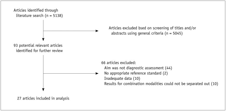
Fig. 1 Flowchart for selection of eligible studies.
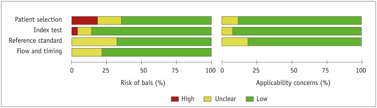
Fig. 2 Assessment of methodological quality of included studies based on Quality Assessment on Diagnostic Accuracy Studies-2 tool.
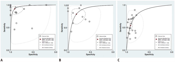
Fig. 3 HSROC curves describing diagnostic accuracy of T2-weighted imging alone for detecting locally recurrent prostate cancer.A. Per-patient analysis of patients who underwent radical prostatectomy. B. Per-patient analysis of patients who underwent radiotherapy. C. Sextant biopsy analysis of patients who underwent radiotherapy. AUC = area under HSROC curve, HSROC = hierarchical summary receiver operating characteristic, SENS = sensitivity, SPEC = specificity
Reference
-
1. Jemal A, Center MM, DeSantis C, Ward EM. Global patterns of cancer incidence and mortality rates and trends. Cancer Epidemiol Biomarkers Prev. 2010; 19:1893–1907. PMID: 20647400.
Article2. Freedland SJ, Presti JC Jr, Amling CL, Kane CJ, Aronson WJ, Dorey F, et al. Time trends in biochemical recurrence after radical prostatectomy: results of the SEARCH database. Urology. 2003; 61:736–741. PMID: 12670557.
Article3. Han M, Partin AW, Zahurak M, Piantadosi S, Epstein JI, Walsh PC. Biochemical (prostate specific antigen) recurrence probability following radical prostatectomy for clinically localized prostate cancer. J Urol. 2003; 169:517–523. PMID: 12544300.
Article4. Rinnab L, Mottaghy FM, Blumstein NM, Reske SN, Hautmann RE, Hohl K, et al. Evaluation of [11C]-choline positron-emission/computed tomography in patients with increasing prostate-specific antigen levels after primary treatment for prostate cancer. BJU Int. 2007; 100:786–793. PMID: 17822459.
Article5. Chism DB, Hanlon AL, Horwitz EM, Feigenberg SJ, Pollack A. A comparison of the single and double factor high-risk models for risk assignment of prostate cancer treated with 3D conformal radiotherapy. Int J Radiat Oncol Biol Phys. 2004; 59:380–385. PMID: 15145151.
Article6. Lee WR, Hanks GE, Hanlon A. Increasing prostate-specific antigen profile following definitive radiation therapy for localized prostate cancer: clinical observations. J Clin Oncol. 1997; 15:230–238. PMID: 8996147.
Article7. Bianco FJ Jr, Scardino PT, Stephenson AJ, Diblasio CJ, Fearn PA, Eastham JA. Long-term oncologic results of salvage radical prostatectomy for locally recurrent prostate cancer after radiotherapy. Int J Radiat Oncol Biol Phys. 2005; 62:448–453. PMID: 15890586.
Article8. Roach M 3rd. The role of PSA in the radiotherapy of prostate cancer. Oncology (Williston Park). 1996; 10:1143–1153. discussion 1154-1161. PMID: 8869957.9. JLuzurier A, Jouve De Guibert PH, Allera A, Feldman SF, Conort P, Simon JM, et al. Dynamic contrast-enhanced imaging in localizing local recurrence of prostate cancer after radiotherapy: limited added value for readers of varying level of experience. J Magn Reson Imaging. 2018; 10. 08. [Epub ahead of print]. DOI: 10.1002/jmri.25991.10. Mazaheri Y, Shukla-Dave A, Goldman DA, Moskowitz CS, Takeda T, Reuter VE, et al. Characterization of prostate cancer with MR spectroscopic imaging and diffusion-weighted imaging at 3 Tesla. Magn Reson Imaging. 2018; 8. 31. pii: S0730-725X(18)30428-4. [Epub ahead of print]. DOI: 10.1016/j.mri.2018.08.025.11. Wu LM, Xu JR, Gu HY, Hua J, Zhu J, Chen J, et al. Role of magnetic resonance imaging in the detection of local prostate cancer recurrence after external beam radiotherapy and radical prostatectomy. Clin Oncol (R Coll Radiol). 2013; 25:252–264. PMID: 23313568.
Article12. Higgins JPT, Green S. Cochrane handbook for systematic reviews of interventions version 5.1.0. The Cochrane Collaboration Web site. 2011. 3. 20. Accessed January 30, 2016. https://training.cochrane.org/handbook.13. Moher D, Liberati A, Tetzlaff J, Altman DG. PRISMA Group. Preferred reporting items for systematic reviews and meta-analyses: the PRISMA statement. Int J Surg. 2010; 8:336–341. PMID: 20171303.
Article14. Whiting PF, Rutjes AW, Westwood ME, Mallett S, Deeks JJ, Reitsma JB, et al. QUADAS-2 Group. QUADAS-2: a revised tool for the quality assessment of diagnostic accuracy studies. Ann Intern Med. 2011; 155:529–536. PMID: 22007046.
Article15. Hollingworth W, Medina LS, Lenkinski RE, Shibata DK, Bernal B, Zurakowski D, et al. Interrater reliability in assessing quality of diagnostic accuracy studies using the QUADAS tool. A preliminary assessment. Acad Radiol. 2006; 13:803–810. PMID: 16777553.16. Chu H, Cole SR. Bivariate meta-analysis of sensitivity and specificity with sparse data: a generalized linear mixed model approach. J Clin Epidemiol. 2006; 59:1331–1332. author reply 1332-1333. PMID: 17098577.
Article17. Wang F, Gatsonis CA. Hierarchical models for ROC curve summary measures: design and analysis of multi-reader, multi-modality studies of medical tests. Stat Med. 2008; 27:243–256. PMID: 17340598.
Article18. Egger M, Smith GD. Bias in location and selection of studies. BMJ. 1998; 316:61–66. PMID: 9451274.19. Harbord RM, Egger M, Sterne JA. A modified test for small-study effects in meta-analyses of controlled trials with binary endpoints. Stat Med. 2006; 25:3443–3457. PMID: 16345038.
Article20. Kim CK, Park BK, Lee HM. Prediction of locally recurrent prostate cancer after radiation therapy: incremental value of 3T diffusion-weighted MRI. J Magn Reson Imaging. 2009; 29:391–397. PMID: 19161194.
Article21. Tamada T, Sone T, Jo Y, Hiratsuka J, Higaki A, Higashi H, et al. Locally recurrent prostate cancer after high-dose-rate brachytherapy: the value of diffusion-weighted imaging, dynamic contrast-enhanced MRI, and T2-weighted imaging in localizing tumors. AJR Am J Roentgenol. 2011; 197:408–414. PMID: 21785087.
Article22. Carbone SF, Pirtoli L, Ricci V, Carfagno T, Tini P, La Penna A, et al. Diffusion-weighted magnetic resonance diagnosis of local recurrences of prostate cancer after radical prostatectomy: preliminary evaluation on twenty-seven cases. Biomed Res Int. 2014; 2014:780816. PMID: 24695416.
Article23. Donati OF, Jung SI, Vargas HA, Gultekin DH, Zheng J, Moskowitz CS, et al. Multiparametric prostate MR imaging with T2-weighted, diffusion-weighted, and dynamic contrast-enhanced sequences: are all pulse sequences necessary to detect locally recurrent prostate cancer after radiation therapy? Radiology. 2013; 268:440–450. PMID: 23481164.
Article24. Kitajima K, Hartman RP, Froemming AT, Hagen CE, Takahashi N, Kawashima A. Detection of local recurrence of prostate cancer after radical prostatectomy using endorectal coil MRI at 3 T: addition of DWI and dynamic contrast enhancement to T2-weighted MRI. AJR Am J Roentgenol. 2015; 205:807–816. PMID: 26397329.
Article25. Hara T, Inoue Y, Satoh T, Ishiyama H, Sakamoto S, Woodhams R, et al. Diffusion-weighted imaging of local recurrent prostate cancer after radiation therapy: comparison with 22-core three-dimensional prostate mapping biopsy. Magn Reson Imaging. 2012; 30:1091–1098. PMID: 22819584.
Article26. Haider MA, Chung P, Sweet J, Toi A, Jhaveri K, Ménard C, et al. Dynamic contrast-enhanced magnetic resonance imaging for localization of recurrent prostate cancer after external beam radiotherapy. Int J Radiat Oncol Biol Phys. 2008; 70:425–430. PMID: 17881141.27. Rouvière O, Valette O, Grivolat S, Colin-Pangaud C, Bouvier R, Chapelon JY, et al. Recurrent prostate cancer after external beam radiotherapy: value of contrast-enhanced dynamic MRI in localizing intraprostatic tumor—correlation with biopsy findings. Urology. 2004; 63:922–927. PMID: 15134982.
Article28. Silverman JM, Krebs TL. MR imaging evaluation with a transrectal surface coil of local recurrence of prostatic cancer in men who have undergone radical prostatectomy. AJR Am J Roentgenol. 1997; 168:379–385. PMID: 9016212.
Article29. Westphalen AC, Kurhanewicz J, Cunha RM, Hsu IC, Kornak J, Zhao S, et al. T2-weighted endorectal magnetic resonance imaging of prostate cancer after external beam radiation therapy. Int Braz J Urol. 2009; 35:171–180. discussion 181-182. PMID: 19409121.
Article30. Morgan VA, Riches SF, Giles S, Dearnaley D, deSouza NM. Diffusion-weighted MRI for locally recurrent prostate cancer after external beam radiotherapy. AJR Am J Roentgenol. 2012; 198:596–602. PMID: 22357998.
Article31. Pucar D, Shukla-Dave A, Hricak H, Moskowitz CS, Kuroiwa K, Olgac S, et al. Prostate cancer: correlation of MR imaging and MR spectroscopy with pathologic findings after radiation therapy-initial experience. Radiology. 2005; 236:545–553. PMID: 15972335.
Article32. Sella T, Schwartz LH, Swindle PW, Onyebuchi CN, Scardino PT, Scher HI, et al. Suspected local recurrence after radical prostatectomy: endorectal coil MR imaging. Radiology. 2004; 231:379–385. PMID: 15064390.
Article33. Sciarra A, Panebianco V, Salciccia S, Osimani M, Lisi D, Ciccariello M, et al. Role of dynamic contrast-enhanced magnetic resonance (MR) imaging and proton MR spectroscopic imaging in the detection of local recurrence after radical prostatectomy for prostate cancer. Eur Urol. 2008; 54:589–600. PMID: 18226441.
Article34. Panebianco V, Sciarra A, Lisi D, Galati F, Buonocore V, Catalano C, et al. Prostate cancer: 1HMRS-DCEMR at 3T versus [(18)F]choline PET/CT in the detection of local prostate cancer recurrence in men with biochemical progression after radical retropubic prostatectomy (RRP). Eur J Radiol. 2012; 81:700–708. PMID: 21330082.
Article35. Cirillo S, Petracchini M, Scotti L, Gallo T, Macera A, Bona MC, et al. Endorectal magnetic resonance imaging at 1.5 Tesla to assess local recurrence following radical prostatectomy using T2-weighted and contrast-enhanced imaging. Eur Radiol. 2009; 19:761–769. PMID: 18825386.
Article36. Casciani E, Polettini E, Carmenini E, Floriani I, Masselli G, Bertini L, et al. Endorectal and dynamic contrast-enhanced MRI for detection of local recurrence after radical prostatectomy. AJR Am J Roentgenol. 2008; 190:1187–1192. PMID: 18430830.
Article37. Linder BJ, Kawashima A, Woodrum DA, Tollefson MK, Karnes J, Davis BJ, et al. Early localization of recurrent prostate cancer after prostatectomy by endorectal coil magnetic resonance imaging. Can J Urol. 2014; 21:7283–7289. PMID: 24978358.38. Wassberg C, Akin O, Vargas HA, Shukla-Dave A, Zhang J, Hricak H. The incremental value of contrast-enhanced MRI in the detection of biopsy-proven local recurrence of prostate cancer after radical prostatectomy: effect of reader experience. AJR Am J Roentgenol. 2012; 199:360–366. PMID: 22826397.
Article39. Boonsirikamchai P, Kaur H, Kuban DA, Jackson E, Hou P, Choi H. Use of maximum slope images generated from dynamic contrast-enhanced MRI to detect locally recurrent prostate carcinoma after prostatectomy: a practical approach. AJR Am J Roentgenol. 2012; 198:W228–W236. PMID: 22358019.
Article40. Westphalen AC, Coakley FV, Roach M 3rd, McCulloch CE, Kurhanewicz J. Locally recurrent prostate cancer after external beam radiation therapy: diagnostic performance of 1.5-T endorectal MR imaging and MR spectroscopic imaging for detection. Radiology. 2010; 256:485–492. PMID: 20551184.
Article41. Coakley FV, Teh HS, Qayyum A, Swanson MG, Lu Y, Roach M 3rd, et al. Endorectal MR imaging and MR spectroscopic imaging for locally recurrent prostate cancer after external beam radiation therapy: preliminary experience. Radiology. 2004; 233:441–448. PMID: 15375223.
Article42. Abd-Alazeez M, Ramachandran N, Dikaios N, Ahmed HU, Emberton M, Kirkham A, et al. Multiparametric MRI for detection of radiorecurrent prostate cancer: added value of apparent diffusion coefficient maps and dynamic contrast-enhanced images. Prostate Cancer Prostatic Dis. 2015; 18:128–136. PMID: 25644248.
Article43. Kim CK, Park BK, Park W, Kim SS. Prostate MR imaging at 3T using a phased-arrayed coil in predicting locally recurrent prostate cancer after radiation therapy: preliminary experience. Abdom Imaging. 2010; 35:246–252. PMID: 19130116.
Article44. Panebianco V, Barchetti F, Sciarra A, Musio D, Forte V, Gentile V, et al. Prostate cancer recurrence after radical prostatectomy: the role of 3-T diffusion imaging in multi-parametric magnetic resonance imaging. Eur Radiol. 2013; 23:1745–1752. PMID: 23377546.
Article45. Kara T, Akata D, Akyol F, Karçaaltıncaba M, Özmen M. The value of dynamic contrast-enhanced MRI in the detection of recurrent prostate cancer after external beam radiotherapy: correlation with transrectal ultrasound and pathological findings. Diagn Interv Radiol. 2011; 17:38–43. PMID: 20703995.46. Drudi FM, Giovagnorio F, Carbone A, Ricci P, Petta S, Cantisani V, et al. Transrectal colour Doppler contrast sonography in the diagnosis of local recurrence after radical prostatectomy--comparison with MRI. Ultraschall Med. 2006; 27:146–151. PMID: 16602039.47. Coakley FV, Hricak H, Wefer AE, Speight JL, Kurhanewicz J, Roach M. Brachytherapy for prostate cancer: endorectal MR imaging of local treatment-related changes. Radiology. 2001; 219:817–821. PMID: 11376276.
Article48. Costello LC, Franklin RB. Concepts of citrate production and secretion by prostate: 2. Hormonal relationships in normal and neoplastic prostate. Prostate. 1991; 19:181–205. PMID: 1946039.
Article49. Zakian KL, Sircar K, Hricak H, Chen HN, Shukla-Dave A, Eberhardt S, et al. Correlation of proton MR spectroscopic imaging with Gleason score based on step-section pathologic analysis after radical prostatectomy. Radiology. 2005; 234:804–814. PMID: 15734935.
Article
- Full Text Links
-
- Actions
-
Cited
- CITED
-
- Close
- Share
-
- Similar articles
-
- Multidisciplinary Functional MR Imaging for Prostate Cancer
- Multiparametric magnetic resonance imaging for prostate cancer: A review and update for urologists
- Medical imaging of prostate cancer
- The Use of Magnetic Resonance Imaging in the Prostate Cancer Primary Diagnostic Pathway: Is It Ready for Primetime?
- Multiparametric MRI in the Detection of Clinically Significant Prostate Cancer
