J Korean Orthop Assoc.
1995 Oct;30(5):1529-1537. 10.4055/jkoa.1995.30.5.1529.
Comparative Analysis of Acromial Morphology in Normal and Impingement Syndrome
- KMID: 2423524
- DOI: http://doi.org/10.4055/jkoa.1995.30.5.1529
Abstract
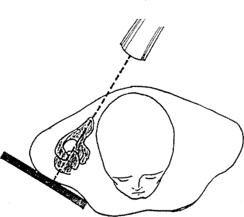
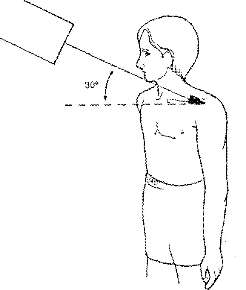
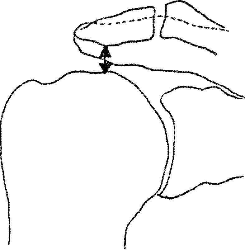
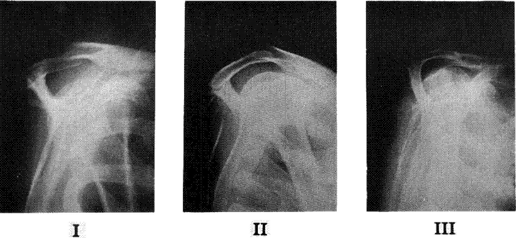
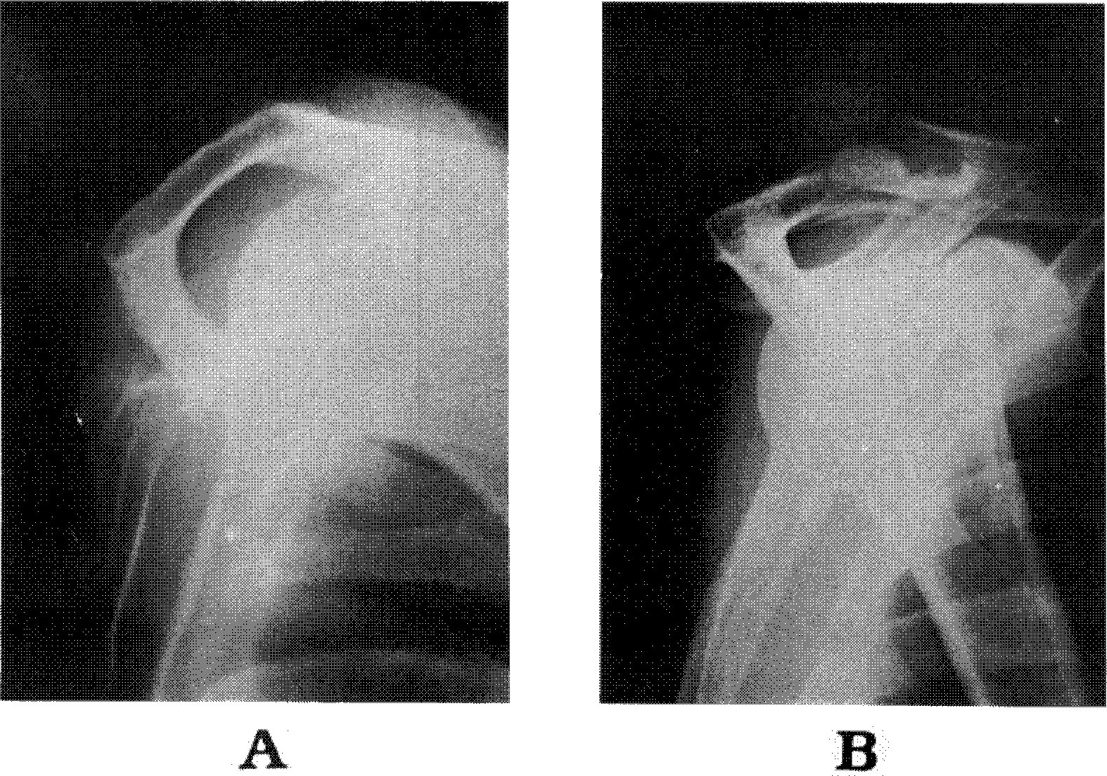
- To identify whether acromial shape, osteophyte, and acromio-humeral interval have effects on impingement syndrome or rotator cuff tear, we reviewed 40 cases of normal group (F:M=22:18), and 30 cases of impingement syndrome(F:M=16:14). Forty cases of normal group aged from 40 to 69 who had no pain, no abrasion sign, no limitation of motion, and normal function of shoulder joint were selected. Thirty cases of impingement syndrome were managed by acromioplasty of direct repair from October, 1993 to May, 1994. Twenty-five cases of 30 were identified rotator cuff tear(RCT), and the others were turned out subacromial abrasion. We reviewed the acromial thickness, the acromial shape, the anterior protuberance, the presence of osteophyte, and the acromio-humeral interval to compare the difference between two groups. Forty-seven point five per cent of normal group had a flat, type I acromion, 47.5% had a curved, type II acromion and 5% were identified by a hooked, type III acromion. However, in subjects with impingement syndrome and RCT, 37% had type I, 20% had type II, and 43% displayed type III. Type III was considerably noticed in the massive tear. In regarding to acromial thickness, normal group had type A(less than 8mm)-37.5%, type B(8-12mm)-62.5%, and the impingement syndrome or RCT group had type A-53%, type B-47%. We couldn't find any significant difference with each group in type III(more than 8mm)-15% in normal, and type I-17%, type II-33%, type III-50% in the impingement syndrome or RCT. It was suggested that the anterior protuberance was related with the evidence of RCT. A-H interval was 10.25mm±1.46mm in normal, and 9.44mm±1.70mm in the impingement syndrome or RCT. There was on significance in A-H interval except rotator cuff arthropathy. Thirty three percent of normal group had osteophytes and 40% of impingement syndrome or RCT had osteophytes on the undersurface of acromion.
Keyword
Figure
Reference
-
1. Bigliani LU, Morrison D, April EW. The morphology of the acromion and its relationship to rotator cuff tears. Orthop Tram. 1986; 10:228–242.2. Bigliani LU, Norris TR, Fisher J. . The relationship between the unfused acromial epiphysis and subacromial impingement lesions. Orthop Trans. 1983; 7((1)):138–154.3. Codman EA. The Shoulder. Rupture of the Supraspinatus Tendon and Other Lesions in or about the Subacromial Bursa. Boston, Thomas Todd. 1934; 67–79.4. Cotton RE, Rideout DF. Tears of The Humeral Rotator Cuff. A Radiological and Pathological Necropsy Survey. J Bone and Joint Surg. 1964; 46-B:314–328.5. Edelson JG, Taitz G. Anatomy of the cora-coacromial arch. Relation to degeneration of the acromion. J Bone and Joint Surg. 1992; 74-B:589–594.
Article6. Flugstad D, Matsen FA, Larry I, Jackins SE. Failed acromioplasty - etiology and prevention. Orthop Trans. 1986; 10:229–245.7. Golding FC. The shoulder-the forgetton joint. British J Radiol,. 1962; 35:149–158.8. Kotzen LM. Roentgen Diagnosis of Rotator Cuff Tear. Report of 48 Surgically Proven Cases. Am J Roentgenol. 1971; 112:507–511.9. Neer CSII. Anterior Aeromioplasty for the Chronic Impingement Syndrome in the Shoulder. A Preliminary Report. J Bone Joint Surg. 1972; 54-A:41–50.10. Neer CSII. impingement Lesions. Clin Orthop. 1983; 173:70–77.
Article11. Neer CSII, Bigliani LU, Hawkins RJ. Rupture of the long head of the biceps related to subacromial impingement. Orthop Trans. 1977; 1:111–130.12. Petersson CJ, Redlund-Johnell I. The Subacromial Space in Normal Shoulder Radiographs. Acta Orthop Scand. 1984; 55:57–58.
Article13. Rockwood CA, Lyons FR. Shoulder impingement syndrome: Diagnosis, Radiographic evaluation and treatment with modified Neer acromioplasty. J Bone Joint Surg:. 75-A:409–424. 1993.14. Snyder SJ, Wuh HCK. A modified classification of the supraspinatus outlet view based on the configuration and the anatomic thickness of the acromion. Orthop Clin N Am. 1993; 24((1)):177–178.15. Weiner DS, Macnab I. Superior Migration of Humeral Head. A Radiological Aid in the Diagnosis of Tears of the Rotator Cuff. J Bone and Joint Surg. 1970; 52-B:524–527.16. Zuckerman J.D., Klummer FJ, Cuomo F., Simon J, Rosenblum S. The influence of the cora-coacromial arch anatomy on rotator cuff tears. J Shoulder and Elbow. 1992; 1:4–14.
- Full Text Links
- Actions
-
Cited
- CITED
-
- Close
- Share
- Similar articles
-
- Study for Acromial Type, Acromial Tilt and Subacromial Distances in Subacromial Impingement Syndrome
- A Study of Acromial Shape, Acromial Angle, Subacromial Distance in Normal Shoulder
- Acromial morphology and morphometry associated with subacromial impingement syndrome
- Acromion Morphology in Coronal and Sagittal Plane; Correlation with Rotator Cuff Syndrome
- Acromial Downslping and Subacromial Interval in Shoulder Impingement Syndrome

