A Rare Case of Subarachnoid Hemorrhage caused by Ruptured Venous Varix Due to Dural Arteriovenous Fistula at the Foramen Magnum Fed Solely by the Ascending Pharyngeal Artery
- Affiliations
-
- 1Department of Neurosurgery, Daegu Fatima Hospital, Daegu, Korea. paulyoonsoolee@hanmail.net
- KMID: 2422606
- DOI: http://doi.org/10.7461/jcen.2018.20.2.120
Abstract
- Dural arteriovenous fistula (D-AVF) at the foramen magnum is an extremely rare disease entity. It produces venous hypertension, and can lead to progressive cervical myelopathy thereafter. On the other hand, the venous hypertension may lead to formation of a venous varix, and it can rarely result in an abrupt onset of subarachnoid hemorrhage (SAH) when the venous varix is ruptured. The diagnosis of D-AVF at the foramen magnum as a cause of SAH may be difficult due to its low incidence. Furthermore, when the D-AVF is fed solely by the ascending pharyngeal artery (APA), it may be missed if the external carotid angiography is not performed. The outcome could be fatal if the fistula is unrecognized. Herein, we report on a rare case of SAH caused by ruptured venous varix due to D-AVF at the foramen magnum fed solely by the APA. A review of relevant literatures is provided, and the treatment modalities and outcomes are also discussed.
Keyword
MeSH Terms
Figure
-
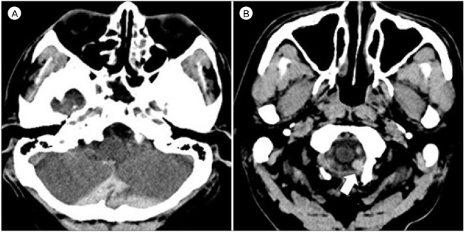
Fig. 1 Initial brain computerized tomography shows subarachnoid hemorrhage at the cistern magna (A). Focal hematoma (white arrow) is observed on the left side of the foramen magnum which is suspected to be the point of rupture (B).
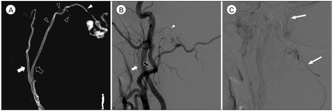
Fig. 2 Three-dimensional reconstruction (A), arterial phase (B), and venous phase (C) of left external carotid angiogram show a dural arteriovenous fistula at the foramen magnum fed by the hypoglossal branch (white arrowheads) of the neuromeningeal trunk (black arrows) of left ascending pharyngeal artery and drained into the suboccipital venous plexus and sigmoid sinus (long white arrows). The pharyngeal trunk (white arrows) and the odontoid arcade (black arrowheads) are also identified. Note the venous varix adjacent to the fistula.
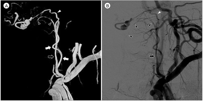
Fig. 3 Three-dimensional reconstruction (A), arterial phase (B) of the left external carotid angiogram at a working angle demonstrate a venous varix distal to the fistula. The hypoglossal branch (white arrowheads) of the neuromeningeal trunk (black arrows) is the only route that leads to the fistulous point. The pharyngeal trunk (white arrows) is overlapped on it. The odontoid arcade (black arrowheads) is also visible.
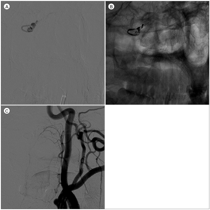
Fig. 4 A superselectvie angiogram clearly shows the configuration of the target (A). Note that the microcatheter should be located as close to the fistulous point as possible. After injecting 0.4 mL of Onyx (B), the fistulous point and the ruptured venous varix are occluded (C).
Reference
-
1. Ansari SA, Lassig JP, Nicol E, Thompson BG, Gemmete JJ, Gandhi D. Transarterial embolization of a cervical dural arteriovenous fistula. Presenting with subarachnoid hemorrhage. Interv Neuroradiol. 2006; 12. 12(4):313–318. PMID: 20569588.2. Aviv RI, Shad A, Tomlinson G, Niemann D, Teddy PJ, Molyneux AJ, et al. Cervical dural arteriovenous fistula manifesting as subarachnoid hemorrhage: report of two cases and literature review. AJNR Am J Neuroradiol. 2004; 5. 25(5):854–858. PMID: 15140735.3. Cavalcanti DD, Reis CV, Hanel R, Abbasi SS, Deshmukh P, Spetzler RF, et al. The ascending pharyngeal artery and its relevance for neurosurgical and endovascular procedures. Neurosurgery. 2009; 12. 65(6 Suppl):114–120. discussion 120. PMID: 19934985.
Article4. Do HM, Jensen ME, Cloft HJ, Kallmes DF, Dion JE. Dural arteriovenous fistula of the cervical spine presenting with subarachnoid hemorrhage. AJNR Am J Neuroradiol. 1999; 2. 20(2):348–350. PMID: 10094368.5. Gaensler EH, Jackson DE Jr, Halbach VV. Arteriovenous fistulas of the cervicomedullary junction as a cause of myelopathy: radiographic findings in two cases. AJNR Am J Neuroradiol. 1990; 5. 11(3):518–521. PMID: 2112318.6. Gross BA, Albuquerque FC, Moon K, Mcdougall CG. The road less traveled: transarterial embolization of dural arteriovenous fistulas via the ascending pharyngeal artery. J NeuroInterv Surg. 2017; 1. 9(1):97–101. PMID: 27581042.
Article7. Guo LM, Zhou HY, Xu JW, Wang GS, Tian X, Wang Y, et al. Dural arteriovenous fistula at the foramen magnum presenting with subarachnoid hemorrhage: case reports and literature review. Eur J Neurol. 2010; 5. 17(5):684–691. PMID: 20050886.
Article8. Hacein-Bey L, Daniels DL, Ulmer JL, Mark LP, Smith MM, Strottmann JM, et al. The ascending pharyngeal artery: branches, anastomoses, and clinical significance. AJNR Am J Neuroradiol. 2002; 8. 23(7):1246–1256. PMID: 12169487.9. Hurst RW, Bagley LJ, Scanlon M, Flamm ES. Dural arteriovenous fistulas of the craniocervical junction. Skull Base Surg. 1999; 9(1):1–7.
Article10. Koch C, Gottschalk S, Giese A. Dural arteriovenous fistula of the lumbar spine presenting with subarachnoid hemorrhage. Case report and review of the literature. J Neurosurg. 2004; 4. 100(4 Suppl Spine):385–391. PMID: 15070151.11. Lee JY, Cho YD, Kwon BJ, Han MH. Dural arteriovenous fistula at the foramen magnum with holocord myelopathy: case report. Neurointervention. 2010; 2. 5(1):53–57.
Article12. Liang G, Gao X, Li Z, Wang X, Zhang H, Wu Z. Endovascular treatment for dural arteriovenous fistula at the foramen magnum: report of five consecutive patients and experience with balloon-augmented transarterial Onyx injection. J Neuroradiol. 2013; 5. 40(2):134–139. PMID: 23433906.
Article13. Mascalchi M, Scazzeri F, Prosetti D, Ferrito G, Salvi F, Quilici N. Dural arteriovenous fistula at the craniocervical junction with perimedullary venous drainage. AJNR Am J Neuroradiol. 1996; Jun-Jul. 17(6):1137–1141. PMID: 8791928.14. Mondel PK, Saraf R, Limaye US. Acute subarachnoid hemorrhage in posterior condylar canal dural arteriovenous fistula: imaging features with endovascular management. J Neurointerv Surg. 2015; 7. 7(7):e26. PMID: 25006042.
Article15. Reinges MHT, Thron A, Mull M, Huffmann BC, Gilsbach JM. Dural arteriovenous fistulae at the foramen magnum. J Neurol. 2001; 3. 248(3):197–203. PMID: 11355153.
Article16. Sadeh-Gonik U, Gory B, Riva R, Labeyrie PE, Signorelli F, Lukaszewicz AC, et al. Ethylene vinyl alcohol copolymer (Onyx®) embolization of cranial dural arteriovenous fistula via the ascending pharyngeal artery. Diagn Interv Imaging. 2016; 6. 97(6):681–685. PMID: 26867991.17. Spiotta AM, Hughes G, Masaryk TJ, Hui FK. Balloon-augmented Onyx embolization of a dural arteriovenous fistula arising from the neuromeningeal trunk of the ascending pharyngeal artery: technical report. J NeuroInterv Surg. 2011; 9. 3(3):300–303. PMID: 21990848.
Article18. Suda S, Katsura KI, Okubo S, Abe A, Kanamaru T, Ueda M, et al. A case of dural arteriovenous fistulas at the craniocervical junction presenting with occipital/neck pain associated with sleep. Intern Med. 2012; 4. 51(8):925–928. PMID: 22504252.
Article19. Sun HS, Yun HS, Song MK, Han JY, Choi IS, Lee SG. Dural arteriovenous fistula on the brain stem and upper cervical spinal cord - a case report -. Ann Rehabil Med. 2011; 10. 35(5):733–737. PMID: 22506199.
Article20. Tanoue S, Goto K, Oota S. Endovascular treatment for dural arteriovenous fistula of the anterior condylar vein with unusual venous drainage: report of two cases. AJNR Am J Neuroradiol. 2005; 9. 26(8):1955–1959. PMID: 16155141.21. Viñuela F, Fox AJ, Pelz DM, Drake CG. Unusual clinical manifestations of dural arteriovenous malformations. J Neurosurg. 1986; 4. 64(4):554–558. PMID: 3950738.
Article22. Willinsky R, Terbrugge K, Lasjaunias P, Montanera W. The variable presentations of craniocervical and cervical dural arteriovenous malformations. Surg Neurol. 1990; 8. 34(2):118–123. PMID: 2367931.
Article23. Wu Q, Wang HD, Shin YS, Zhang X. Brainstem congestion due to dural arteriovenous fistula at the craniocervical junction. J Korean Neurosurg Soc. 2014; 3. 55(3):152–155. PMID: 24851151.24. Zhao J, Xu F, Ren J, Manjila S, Bambakidis NC. Dural arteriovenous fistulas at the craniocervical junction : a systematic review. J NeuroInterv Surg. 2016; 6. 8(6):648–653. PMID: 26041099.
- Full Text Links
-
- Actions
-
Cited
- CITED
-
- Close
- Share
-
- Similar articles
-
- Dural Arteriovenous Fistula of Jugular Foramen with Subarachnoid Hemorrhage : Selective Transarterial Embolization
- Dural Arteriovenous Fistula at the Foramen Magnum with Holocord Myelopathy: Case Report
- Traumatic Intracerebral and Subarachnoid Hemorrhage Due to a Ruptured Pseudoaneurysm of Middle Meningeal Artery Accompanied by a Medial Sphenoid Wing Dural Arteriovenous Fistula
- Borden Type I Sigmoid Sinus Dural Arteriovenous Fistula Presenting as Subarachnoid Hemorrhage from a Feeding Artery Aneurysm of the Anterior Inferior Cerebellar Artery: A Case Report
- Intracranial Dural Arteriovenous Fistula Draining into Spinal Perimedullary Veins: A Rare Cause of Myelopathy
