Evaluation of the Accuracy in Maximum Intensity Projection Images of Cerebral Computed Tomographic Angiography for the Diagnosis of Cerebral Vasospasm Following Subarachnoid Hemorrhage, in Comparison to Digital Subtraction Angiography
- Affiliations
-
- 1Department of Neurosurgery, Yeungnam University Medical Center, Daegu, Korea. jyjns@yu.ac.kr
- 2Department of Radiology, Yeungnam University Medical Center, Daegu, Korea.
- KMID: 2422554
- DOI: http://doi.org/10.7461/jcen.2018.20.1.5
Abstract
OBJECTIVE
The purpose of this retrospective study is to determine the accuracy of maximum intensity projection (MIP) images of computed tomographic angiography (CTA) for diagnosis of cerebral vasospasm (CV) following subarachnoid hemorrhage (SAH) compared with that of digital subtraction angiography (DSA).
MATERIALS AND METHODS
For patients admitted to our hospital for SAH, MIP images of CTA and DSA were checked at admission, and images were taken again 1 week later. This protocol was used in 39 cases. MIP images of CTA and DSA examinations were reviewed by two independent readers.
RESULTS
Accuracy of MIP images of CTA in various arterial segments, using DSA as the gold standard: the sensitivity, specificity, positive predictive value (PPV), negative predictive value (NPV), and accuracy for different segments varied from 84 to 97, 33-100, 84-100%, 25-85, and 79-97%, respectively, for readers. Accuracy of CTA in various vasospasm severity, using DSA as the gold standard: the sensitivity, specificity, PPV, NPV, and accuracy for different vasospasm severity varied from 44 to 100, 69-100, 36-100%, 61-100, and 88-100%, respectively, for readers. Accuracy of CTA in central segments versus peripheral segments, using DSA as the gold standard: the sensitivity, specificity, PPV, NPV, and accuracy for central segments and peripheral segments varied from 90 to 94, 68-83, 93-97%, 56-69, and 87-93%, respectively, for readers.
CONCLUSION
MIP imaging of CTA is a useful modality when diagnosing CV after SAH.
Keyword
MeSH Terms
Figure
-
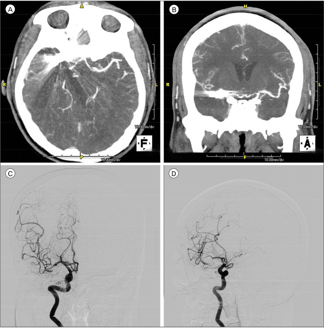
Fig. 1 (A) Right MCA was not visible in the CTA due to the coil artifact. (B) Right MCA was not visible in the CTA due to the coil artifact. (C) Right MCA was clearly confirmed in the DSA regardless of the coil. (D) Right MCA was clearly confirmed in the DSA regardless of the coil. MCA = middle cerebral artery; CTA = computed tomographic angiography; DSA = digital subtraction angiography.
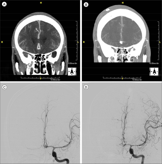
Fig. 2 (A) Both ACA are normal with no vasospasms shown in the initial CTA. (B) Both ACA showed severe vasospasms in the follow-up CTA. (C) Both ACA are normal with no vasospasms shown in the initial DSA. (D) Both ACA showed severe vasospasms in the follow-up DSA. ACA = anterior cerebral artery; CTA = computed tomographic angiography; DSA = digital subtraction angiography.
Reference
-
1. Anderson GB, Ashforth R, Steinke DE, Findlay JM. CT angiography for the detection of cerebral vasospasm in patients with acute subarachnoid hemorrhage. AJNR Am J Neuroradiol. 2000; Jun-Jul. 21(6):1011–1015. PMID: 10871004.2. Chaudhary SR, Ko N, Dillon WP, Yu MB, Liu S, Criqui GI, et al. Prospective evaluation of multidetector-row CT angiography for the diagnosis of vasospasm following subarachnoid hemorrhage: a comparison with digital subtraction angiography. Cerebrovasc Dis. 2008; 25(1-2):144–150. PMID: 18073468.
Article3. Cloft HJ, Joseph GJ, Dion JE. Risk of cerebral angiography in patients with subarachnoid hemorrhage, cerebral aneurysm, and arteriovenous malformation: a meta-analysis. Stroke. 1999; 2. 30(2):317–320. PMID: 9933266.4. Dorsch NW, Young N, Kingston RJ, Compton JS. Early experience with spiral CT in the diagnosis of intracranial aneurysms. Neurosurgery. 1995; 1. 36(1):230–236. discussion 236-8. PMID: 7708163.
Article5. Earnest F 4th, Forbes G, Sandok BA, Piepgras DG, Faust RJ, Ilstrup DM, et al. Complications of cerebral angiography: prospective assessment of risk. AJR Am J Roentgenol. 1984; 2. 142(2):247–253. PMID: 6198889.
Article6. Ferguson SD, Rosen DS, Bardo D, Macdonald RL. Arterial diameters on catheter and computed tomographic angiography. World Neurosurg. 2010; 3. 73(3):165–173. discussion e25. PMID: 20860954.
Article7. Heiserman JE, Dean BL, Hodak JA, Flom RA, Bird CR, Drayer BP, et al. Neurologic complications of cerebral angiography. AJNR Am J Neuroradiol. 1994; 9. 15(8):1401–1407. discussion 1408-11. PMID: 7985557.8. Levi CR, O'Malley HM, Fell G, Roberts AK, Hoare MC, Royle JP, et al. Transcranial Doppler detected cerebral microembolism following carotid endarterectomy. High microembolic signal loads predict postoperative cerebral ischaemia. Brain. 1997; 4. 120(Pt 4):621–629. PMID: 9153124.
Article9. Okada Y, Shima T, Nishida M, Yamane K, Hatayama T, Yamanaka C, et al. Comparison of transcranial Doppler investigation of aneurysmal vasospasm with digital subtraction angiographic and clinical findings. Neurosurgery. 1999; 9. 45(3):443–449. discussion 449-50. PMID: 10493365.
Article10. Otawara Y, Ogasawara K, Ogawa A, Sasaki M, Takahashi K. Evaluation of vasospasm after subarachnoid hemorrhage by use of multislice computed tomographic angiography. Neurosurgery. 2002; 10. 51(4):939–942. discussion 942-3. PMID: 12234400.
Article11. Pryor JC, Setton A, Nelson PK, Berenstein A. Complications of diagnostic cerebral angiography and tips on avoidance. Neuroimaging Clin N Am. 1996; 8. 6(3):751–758. PMID: 8873102.12. Shankar JJ, Tan IY, Krings T, Terbrugge K, Agid R. CT angiography for evaluation of cerebral vasospasm following acute subarachnoid haemorrhage. Neuroradiology. 2012; 3. 54(3):197–203. PMID: 21541687.
Article13. Sloan MA, Haley EC Jr, Kassell NF, Henry ML, Stewart SR, Beskin RR, et al. Sensitivity and specificity of transcranial Doppler ultrasonography in the diagnosis of vasospasm following subarachnoid hemorrhage. Neurology. 1989; 11. 39(11):1514–1518. PMID: 2682350.
Article14. Takagi R, Hayashi H, Kobayashi H, Kumazaki T, Isayama K, Ikeda Y, et al. Three-dimensional CT angiography of intracranial vasospasm following subarachnoid haemorrhage. Neuroradiology. 1998; 10. 40(10):631–635. PMID: 9833891.
Article15. Tamatani S, Sasaki O, Takeuchi S, Fujii Y, Koike T, Tanaka R. Detection of delayed cerebral vasospasm, after rupture of intracranial aneurysms, by magnetic resonance angiography. Neurosurgery. 1997; 4. 40(4):748–753. discussion 753-4. PMID: 9092848.
Article16. Treggiari-Venzi MM, Suter PM, Romand JA. Review of medical prevention of vasospasm after aneurysmal subarachnoid hemorrhage: a problem of neurointensive care. Neurosurgery. 2001; 2. 48(2):249–261. discussion 261-2. PMID: 11220367.
Article17. Vernieri F, Pasqualetti P, Passarelli F, Rossini PM, Silvestrini M. Outcome of carotid artery occlusion is predicted by cerebrovascular reactivity. Stroke. 1999; 3. 30(3):593–598. PMID: 10066857.
Article18. Waugh JR, Sacharias N. Arteriographic complications in the DSA era. Radiology. 1992; 1. 182(1):243–246. PMID: 1727290.
Article19. Wintermark M, Ko NU, Smith WS, Liu S, Higashida RT, Dillon WP. Vasospasm after subarachnoid hemorrhage: utility of perfusion CT and CT angiography on diagnosis and management. AJNR Am J Neuroradiol. 2006; 1. 27(1):26–34. PMID: 16418351.20. Yoon DY, Choi CS, Kim KH, Cho BM. Multidetector-row CT angiography of cerebral vasospasm after aneurysmal subarachnoid hemorrhage: comparison of volume-rendered images and digital subtraction angiography. AJNR Am J Neuroradiol. 2006; 2. 27(2):370–377. PMID: 16484413.
- Full Text Links
-
- Actions
-
Cited
- CITED
-
- Close
- Share
-
- Similar articles
-
- Multidetector-Row CT Angiography of Cerebral Vasospasm after Aneurysmal Subarachnoid Hemorrhage: Comparison of Bone Subtraction and Standard CT Angiography with Digital Subtraction Angiography
- Role of Multislice Computerized Tomographic Angiography in Vasospasm Following Aneurysmal Subarachnoid Hemorrhage
- Cerebral Venous Thrombosis Presenting as Isolated Subarachnoid Hemorrhage
- Evaluation of Subarachnoid Hemorrhage due to Aneurysmal Rupture and Cerebral Vasospasm by CT
- Usefulness of Computed Tomographic Angiography(CTA) in the Evaluation of Cerebral Aneurysms
