Postoperative Hemorrhagic Occlusive Retinal Vasculitis with Intracameral Vancomycin
- Affiliations
-
- 1Department of Ophthalmology, Jeju National University Hospital, Jeju National University School of Medicine, Jeju, Korea. muse1016@naver.com forgotten100@gmail.com
- KMID: 2422076
- DOI: http://doi.org/10.3341/kjo.2018.0043
Abstract
- No abstract available.
Figure
-
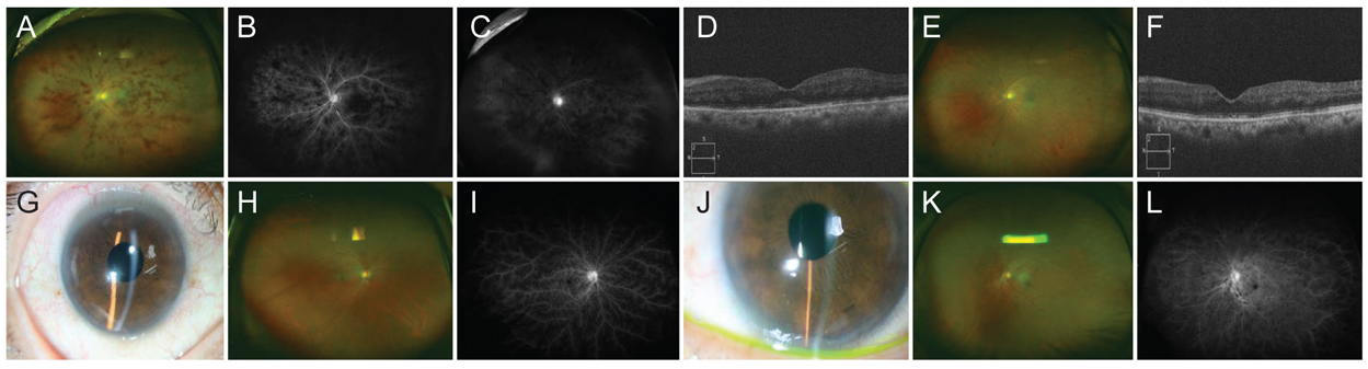
Fig. 1 Wide-field fundus photograph, wide-field fluorescein angiograph, optical coherence tomography and anterior segment photograph images of (A–F) case 1, (G–I) case 2 right eye, and (J–L) case 2 left eye. (A) Wide-field fundus photograph showing multiple patch patterned retinal hemorrhages along venules. The retinal veins are not tortuous or dilated. (B) Wide-field fluorescein angiograph showing multiple vessel occlusion with non-perfusion area (early flame angiogram). (C) Wide-field fluorescein angiograph demonstrating diffuse retinal perivascular leakage and cuffing. (D) Optical coherence tomography showing macular edema with subretinal fluid. (E) Wide-field fundus photograph showing the improved retinal hemorrhages after 8 months of treatment with intravitreal bevacizumab, topical and systemic corticosteroid. (F) Optical coherence tomography showing the absorbed subretinal fluid in the right eye. (G) Anterior segment photograph of the right eye demonstrating iris neovascularization. (H) Wide-field fundus photograph showing the multiple patch patterned retinal hemorrhages. The retinal veins are not tortuous or dilated. (I) Wide-field fluorescein angiograph demonstrating perivascular leakage and peripheral vascular occlusion with extended area of non-perfusion. (J) Anterior segment photograph of the left eye showing the severe neovascularization of iris at the inferonasal aspect of the iris. (K) Wide-field fundus photograph showing multiple scattered intraretinal and perivascular hemorrhages occurred faster than the fellow eye. The retinal veins are not tortuous or dilated. (L) Wide-field fluorescein angiograph showing perivascular leakage with non-perfusion areas in the peripheral retina.
Reference
-
1. Hatch WV, Cernat G, Wong D, et al. Risk factors for acute endophthalmitis after cataract surgery: a population-based study. Ophthalmology. 2009; 116:425–430.
Article2. Chang DF, Braga-Mele R, Henderson BA, et al. Antibiotic prophylaxis of postoperative endophthalmitis after cataract surgery: results of the 2014 ASCRS member survey. J Cataract Refract Surg. 2015; 41:1300–1305.
Article3. Nicholson LB, Kim BT, Jardon J, et al. Severe bilateral ischemic retinal vasculitis following cataract surgery. Ophthalmic Surg Lasers Imaging Retina. 2014; 45:338–342.
Article4. Witkin AJ, Shah AR, Engstrom RE, et al. Postoperative hemorrhagic occlusive retinal vasculitis: expanding the clinical spectrum and possible association with vancomycin. Ophthalmology. 2015; 122:1438–1451.5. Witkin AJ, Chang DF, Jumper JM, et al. Vancomycin-associated hemorrhagic occlusive retinal vasculitis: clinical characteristics of 36 eyes. Ophthalmology. 2017; 124:583–595.
- Full Text Links
-
- Actions
-
Cited
- CITED
-
- Close
- Share
-
- Similar articles
-
- A Case of Acute Multifocal Hemorrhagic Retinal Vasculitis
- Retinal Occlusive Vasculitis after Intravitreal Injection of Brolucizumab
- A Case of Severe Retinal Vaso-Occlusive Disease in Systemic Lupus Erythematosus
- A Case of Juvenile Dermatomyositis Accompanied by Hemorrhagic Retinal Vasculitis and Multiple Gastrointestinal Bleeding
- Prophylactic Intracameral Vancomycin Irrigation and Cystoid Macular Edema
