Introduction of a New Skeletal Imaging Instrument: The Low Radiating-Dose EOS System
- Affiliations
-
- 1Department of Orthopedic Surgery, Konyang University College of Medicine, Daejeon, Korea. ktk1113@kyuh.ac.kr
- KMID: 2421614
- DOI: http://doi.org/10.4184/jkss.2018.25.2.74
Abstract
- STUDY DESIGN: Literature review.
OBJECTIVE
The aim of this article is to introduce the EOS imaging device, with a focus on spinal and pelvic alignment. SUMMARY OF LITERATURE REVIEW: The EOS imaging device can obtain images of spinal and pelvic alignment with almost no distortion, using a low radiation dose.
MATERIALS AND METHODS
We searched for studies related to the use of EOS imaging device for spinal and pelvic alignment.
RESULTS
The EOS is not only capable of simultaneously obtaining paired anteroposterior and lateral X-ray images with a low radiation dose, but also can reconstruct the image as if it was acquired in the patient's reference plane, limiting the distortion to the patient's thickness instead of the whole distance between the source and detector. The EOS device also has the advantage of accurately measuring the sagittal alignment of the spine and pelvis and the torsional deformity of the lower limbs, as the subject can be imaged while standing upright in a weight-bearing posture.
CONCLUSIONS
EOS is a new diagnostic technique that can detect spinal and pelvic alignment and deformities of the lower limbs under weight-bearing conditions with a low radiation dose.
Keyword
Figure
-
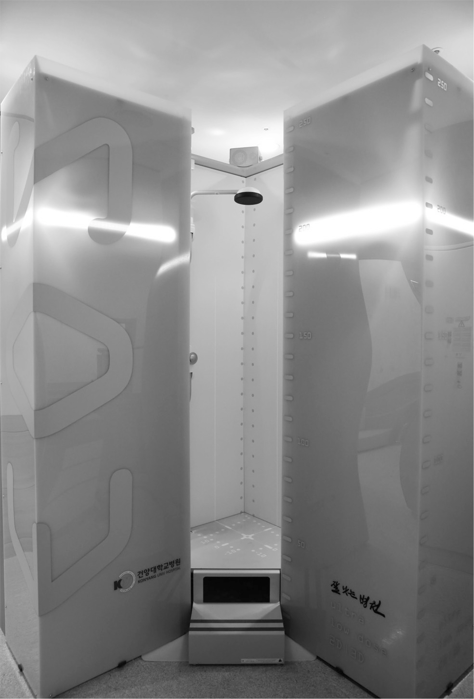
Fig 1. EOS is an open rectangular cage that is 2 m wide and 2.7 m tall.
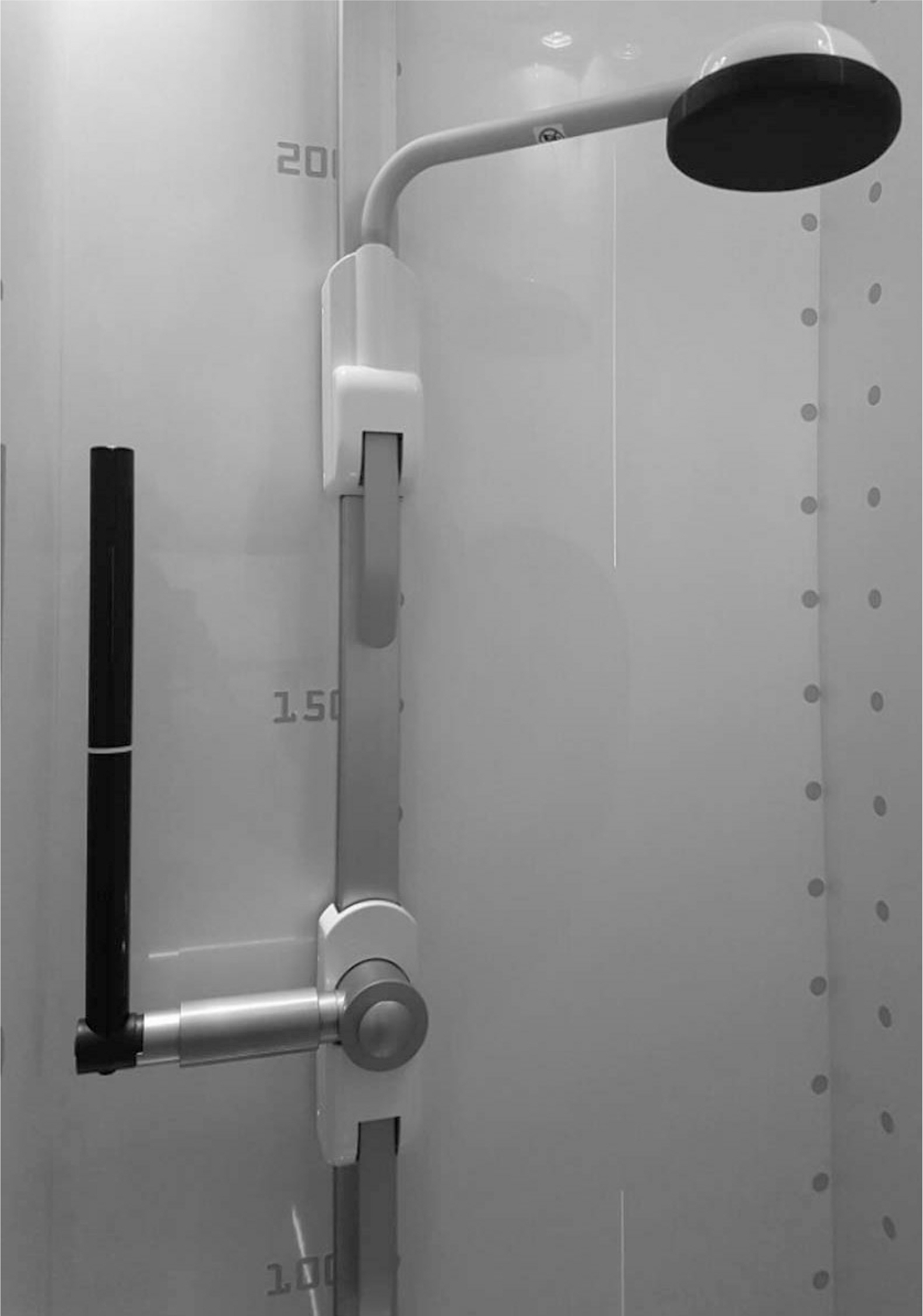
Fig 2. An X-ray tube and a sensor installed at a 90° angle, taking images from all sides at the same time, and a safety bar and a device fixing the patient's head in place while the image is obtained.
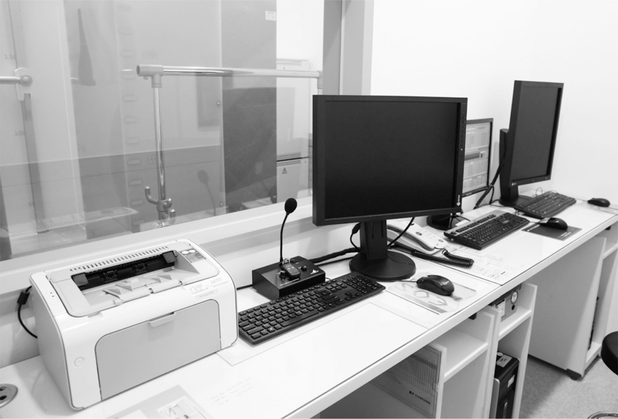
Fig 3. A separated EOS station protected from the X-ray. The length and angles of the obtained images are measured and reproduced as 3-dimensional images by the EOS software at this station.

Fig 4. Diagrammatic representation of the amplification of the low-dose primary X-ray beam through the Charpak chamber.

Fig 5. EOS scans the patient's anteroposterior and lateral sides simultaneously while the patient has both hands placed on the maxillary sinus under the eyes and has paused after inhaling in a weight-bearing position.
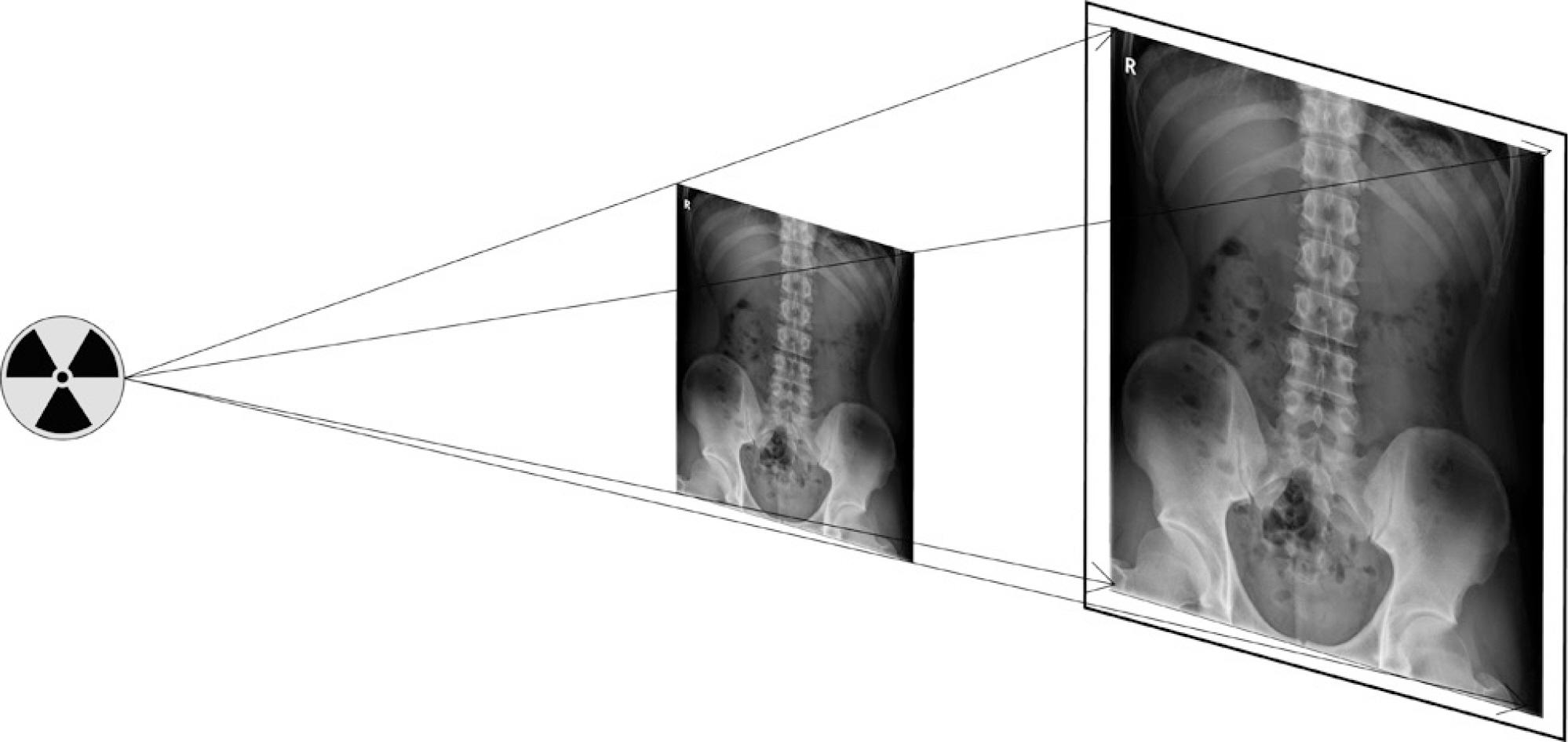
Fig 6. Distortion caused by the conical projection from the center to the edges of the radiograph, increasing the scale of error for structures farther from the central region.

Fig 7. The cylindrical projection image corrected by the system's visualization interface, which limits distortion regardless of the patient's thickness and the distance between the object and the detector.

Fig 8. When a landmark is indicated on 2-dimensional images of the anteroposterior and lateral planes of the spine, the EOS imaging system provides various measures.
Reference
-
1. Deschenes S, Charron G, Beaudoin G, et al. Diagnostic imaging of spinal deformities: reducing patients radiation dose with a new slot-scanning X-ray imager. Spine. 2010 Apr 20; 35(9):989–94. DOI: 10.1097/brs.0b013e3181bdcaa4.2. Escott BG, Ravi B, Weathermon AC, et al. EOS Low-Dose Radiography: A Reliable and Accurate Upright Assessment of Lower-Limb Lengths. J Bone Joint Surg Am. 2013 Dec 4; 95(23):E831–7. DOI: 10.2106/jbjs.l.00989.3. Gurney B. Leg length discrepancy. Gait posture. 2002 Apr; 15(2):195–206. DOI: 10.1016/s0966-6362 (01)00148-5.
Article4. Guichet JM, Spivak JM, Trouilloud P, et al. Lower limb-length discrepancy. An epidemiologic study. Clin Orthop Relat Res. 1991 Nov; 272:235–41. DOI: 10.1097/00003086-199111000-00035.5. Melhem E, Assi A, Rachkidi RE, et al. EOS biplanar X-ray imaging: concept, developments, benefits, and limitations. J Child Orthop. 2016 Feb 16; 10(1):1–14. DOI: 10.1007/s11832-016-0713-0.
Article6. Humbert L, De Guise JA, Aubert B, et al. 3D reconstruction of the spine from biplanar X-rays using parametric models based on transversal and longitudinal inferences. Med Eng Phys. 2009 Jul; 31(6):681–7. DOI: 10.1016/j.medengphy.2009.01.003.
Article7. Chaibi Y, Cresson T, Aubert B, et al. Fast 3D reconstruction of the lower limb using a parametric model and statistical inferences and clinical measurements calculation from biplanar X-rays. Comput Methods Biomech Biomed Engin. 2012 May; 15(5):457–66. DOI: 10.1080/10255842.2010.540758.
Article8. Laporte S, Skalli W, De Guise JA, et al. A biplanar reconstruction method based on 2D and 3D contours: application to the distal femur. Comput Methods Biomech Biomed Engin. 2003 Feb; 6(1):1–6. DOI: 10.1080/1025584031000065956.
Article9. Le Bras A, Laporte S, Mitton D, et al. Three-Dimensional (3D) detailed reconstruction of human vertebrae from low-dose digital stereoradiography. Eur J Orthop Surg Traumatol. 2003 Jun; 13(2):57–62. DOI: 10.1007/s00590-003-0074-5.
Article10. Mitton D, Zhao K, Bertrand S, et al. 3D reconstruction of the ribs from lateral and frontal X-rays in comparison to 3D CT-scan reconstruction. J Biomech. 2008; 41(3):706–10. DOI: 10.1016/j.jbiomech.2007.09.034.
Article11. Rousseau M-A, Laporte S, Chavary E, et al. Reproducibility of measuring the shape and three-dimensional position of cervical vertebrae in upright position using the EOS stereoradiography system. Spine. 2007 Nov; 32(23):2569–72. DOI: 10.1097/brs.0b013e318158cba2.
Article12. Somoskeo S, Tunyogi-Csapo M, Bogyo C, et al. Accuracy and reliability of coronal and sagittal spinal curvature data based on patient-specific three-dimensional models created by the EOS 2D/3D imaging system. Spine J. 2012 Nov; 12(11):1052–9. DOI: 10.1016/j.spinee.2012.10.002.13. Lin B, Zhang WB, Cai TY, et al. Analysis of sagittal balance using spinopelvic parameters in ankylosing spondylitis patients treated with vertebral column decancellation surgery. Acta Orthop Belg. 2015 Sep; 81(3):538–45. DOI: 10.1097/bsd.0b013e31829186c1.14. Jiang L, Qiu Y, Xu L, et al. Sagittal spinopelvic alignment in adolescents associated with Scheuermann's kyphosis: a comparison with normal population. Eur Spine J. 2014 Mar 18; 23(7):1420–6. DOI: 10.1007/s00586-014-3266-2.
Article15. Dong YL, Zhao H, Zhang JG, et al. Effects of pedicle subtraction osteotomy on spinopelvic parameters of ankylosing spondylitis. Zhonghua Yi Xue Za Zhi. 2013; 93:1138–41. DOI: doi.org/10.1007/s00586-009-1158-7.16. Roussouly P, Pinheiro-Franco JL. Biomechanical analysis of the spino-pelvic organization and adaptation in pathology. Eur Spine J. 2011 Sep; 20(5 Suppl):609–18. DOI: 10.1007/s00586-011-1928-x.
Article17. Rose PS, Bridwell KH, Lenke LG, et al. Role of pelvic incidence, thoracic kyphosis, and patient factors on sagittal plane correction following pedicle subtraction osteotomy. Spine. 2009 Apr 15; 34:785–91. DOI: 10.1097/brs.0b013e31819d0c86.
Article18. Bourghli A, Aunoble S, Reebye O, et al. Correlation of clinical outcome and spinopelvic sagittal alignment after surgical treatment of low-grade isthmic spondylolisthesis. Eur Spine J. 2011 Aug 2; 20(5 Suppl):663–8. DOI: 10.1007/s00586-011-1934-z.
Article19. Schwab F, Lafage V, Patel A, et al. Sagittal plane considerations and the pelvis in the adult patient. Spine. 2009 Aug; 34(17):1828–33. DOI: 10.1097/brs.0b013e3181a13c08.
Article20. Guenoun B, Zadegan F, Aim F, et al. Reliability of a new method for lower-extremity measurements based on stereoradiographic three-dimensional reconstruction. Orthop Traumatol Surg Res. 2012 Sep; 98(5):506–13. DOI: 10.1016/j.otsr.2012.03.014.
Article21. Lazenaec JY, Rousseau MA, Rangel A, et al. Orientations du pelvis et de la piece acetabulaire des protheses totales de hanche en position assise et debout: reproductibilite des mesures sulvant le systeme rediographique utilise. EOS ou conventionnel. Rev Chir Orhop. 2011 Jun; 97(4):375–83. DOI: 10.1016/j.rcot.2011.03.036.22. Folinais D, Thelen P, Delin C, et al. Measuring femoral and rotational alignment: EOS system versus computed tomography. Orthop Traumatol Surg Res. 2013 Sep; 99(5):509–16. DOI: 10.1016/j.otsr.2012.12.023.
Article
- Full Text Links
-
- Actions
-
Cited
- CITED
-
- Close
- Share
-
- Similar articles
-
- Sagittal Parameters of Spine and Pelvis in Young Adults Using the EOS Imaging System: Prospective Study of 92 Asymptomatic Subjects
- Reliability of the EOS Imaging System for Assessment of the Spinal and Pelvic Alignment in the Sagittal Plane
- Medical genomic approach to early-onset scoliosis
- Comparison of Whole Spine Sagittal Alignment in Patients with Spinal Disease between EOS Imaging System versus Conventional Whole Spine X-ray
- Thymol Rich Thymbra capitata Essential Oil Inhibits Quorum Sensing, Virulence and Biofilm Formation of Beta Lactamase Producing Pseudomonas aeruginosa
