Coexistence of Intracranial Squamous Cell Carcinoma and Epidermoid Cyst: a Case with Consecutive Imaging Findings
- Affiliations
-
- 1Department of Radiology and Research Institute of Radiology, University of Ulsan College of Medicine, Seoul, Korea. jieunp@gmail.com
- KMID: 2421549
- DOI: http://doi.org/10.13104/imri.2018.22.3.172
Abstract
- In contrast to well-known imaging findings of intracranial epidermoid cysts on magnetic resonance imaging, those of intracranial squamous cell carcinoma (SqCC) are relatively unknown. We present a case of coexistence of intracranial SqCC and epidermoid cyst, with consecutive follow up over 14 months. Based on our case, a solid enhancing portion adjacent to a typically-looking epidermoid cyst may become a clue for coexistence of intracranial SqCC. An initial contrast enhancement and/or heterogeneous signal on diffusion weighted imaging may become a useful diagnostic clue, but more importantly, sudden rapid growth is important in formulating diagnosis.
MeSH Terms
Figure
-
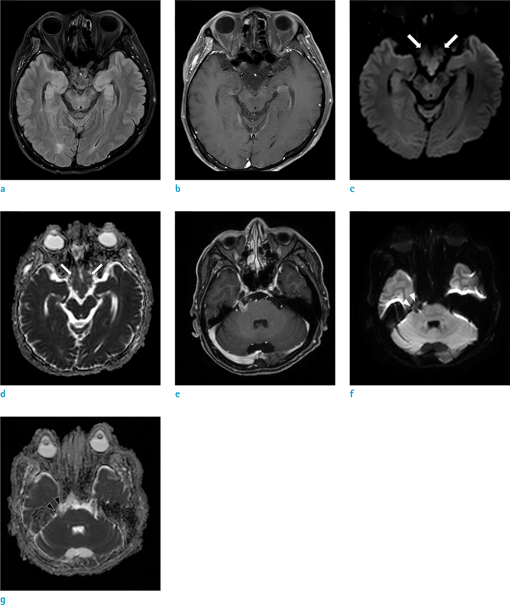
Fig. 1 Initial MRIs were taken at a local hospital, which show lobulated lesion in the basal cistern on (a) FLAIR image, and (b) contrast-enhanced T1WI shows no definite enhancing portion in the lesion. (c) DWI and (d) ADC images show diffusion restriction of the lesion (arrows). Another 1.3 cm sized enhancing lesion was noted in the right CP angle on (e) contrast-enhanced T1WI. Mild but heterogeneous diffusion restriction is shown on (f) DWI and (g) ADC (arrowheads). ADC = apparent diffusion coefficient; DWI = diffusion-weighted image; FLAIR = fluid-attenuated inversion recovery; MRI = magnetic resonance imaging; T1WI = T1-weighted image
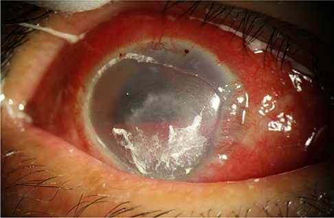
Fig. 2 Ophthalmologic examination and clinical photo show severe corneal ulceration on the right side, which was getting worse despite proper management.
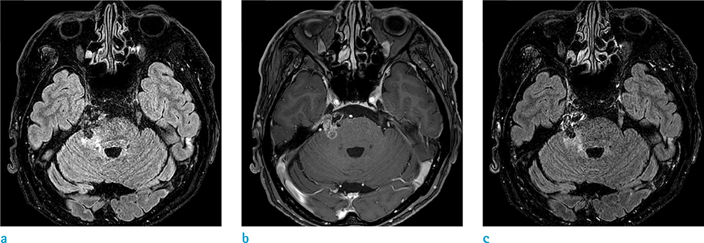
Fig. 3 Second MRIs were taken 5 months after the initial MRIs. The high signal intensity lesion on (a) FLAIR image is more prominent since the previous examination, and the enhancing lesion on (b) T1WI has increased in size as well, with minimal enhancement on (c) contrast-enhanced FLAIR image.
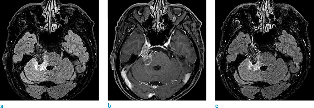
Fig. 4 Third MRIs were taken 9 months after the second MRIs or 14 months after the initial MRIs. The extent of high signal intensity lesion on (a) FLAIR image has been increased, and the size of enhancing lesion on (b) T1WI increased further, with minimal enhancement on (c) contrast-enhanced FLAIR image.
Reference
-
1. Ahmed I, Auguste KI, Vachhrajani S, Dirks PB, Drake JM, Rutka JT. Neurosurgical management of intracranial epidermoid tumors in children. Clinical article. J Neurosurg Pediatr. 2009; 4:91–96.2. Roh TH, Park YS, Park YG, Kim SH, Chang JH. Intracranial squamous cell carcinoma arising in a cerebellopontine angle epidermoid cyst: a case report and literature review. Medicine (Baltimore). 2017; 96:e9423.3. Wagle WA, Jaufmann B, Mincy JE. Magnetic resonance imaging of fourth ventricular epidermoid tumors. Arch Neurol. 1991; 48:438–440.
Article4. Kim MS, Kim OL. Primary intracranial squamous cell carcinoma in the brain stem with a cerebellopontine angle epidermoid cyst. J Korean Neurosurg Soc. 2008; 44:401–404.
Article5. Hamlat A, Hua ZF, Saikali S, et al. Malignant transformation of intra-cranial epithelial cysts: systematic article review. J Neurooncol. 2005; 74:187–194.
Article6. Uchino A, Hasuo K, Matsumoto S, et al. Intracranial epidermoid carcinoma: CT and MRI. Neuroradiology. 1995; 37:155–158.
Article7. Abramson RC, Morawetz RB, Schlitt M. Multiple complications from an intracranial epidermoid cyst: case report and literature review. Neurosurgery. 1989; 24:574–578.
Article8. Nakao Y, Nonaka S, Yamamoto T, et al. Malignant transformation 20 years after partial removal of intracranial epidermoid cyst--case report. Neurol Med Chir (Tokyo). 2010; 50:236–239.9. Ozutemiz C, Ada E, Ersen A, Ozer E. Imaging findings of an epidermoid cyst with malignant transformation to squamous cell carcinoma. Turk Neurosurg. 2017; 27:312–315.
Article10. Nawashiro H, Higo R, Tokumaru AM, Tsuzuki N, Shima K. Diffusion-weighted MRI of an intracranial epidermoid with malignant transformation. Neuroradiology. 2001; 43:891.
Article
- Full Text Links
-
- Actions
-
Cited
- CITED
-
- Close
- Share
-
- Similar articles
-
- Primary Intracranial Epidermoid Carcinoma
- Primary Intracranial Squamous Cell Carcinoma in the Brain Stem with a Cerebellopontine Angle Epidermoid Cyst
- Malignant Transformation of an Epidermoid Cyst in the Cerebellopontine Angle
- Squamous cell carcinoma arising from a long-standing epidermoid cyst of the back
- Epidermoid Cyst after Groin Flap Mimicking Malignancy
