CD57 (Leu-7, HNK-1) immunoreactivity seen in thin arteries in the human fetal lung
- Affiliations
-
- 1Department of Anatomy, Tokyo Dental College, Tokyo, Japan. yamamotomasahito@tdc.ac.jp
- 2Department of Anatomy, Wuxi Medical School, Jiangnan University, Wuxi, China.
- 3Division of Internal Medicine, Iwamizawa Asuka Hospital, Iwamizawa, Japan.
- 4Department of Dentistry, Jikei University School of Medicine, Tokyo, Japan.
- KMID: 2421174
- DOI: http://doi.org/10.5115/acb.2018.51.2.105
Abstract
- CD57 (synonyms: Leu-7, HNK-1) is a well-known marker of nerve elements including the conductive system of the heart, as well as natural killer cells. In lung specimens from 12 human fetuses at 10-34 weeks of gestation, we have found incidentally that segmental, subsegmental, and more peripheral arteries strongly expressed CD57. Capillaries near developing alveoli were often or sometimes positive. The CD57-positive tissue elements within intrapulmonary arteries seemed to be the endothelium, internal elastic lamina, and smooth muscle layer, which corresponded to tissue positive for a DAKO antibody reactive with smooth muscle actin we used. However, the lobar artery and pulmonary arterial trunk as well as bronchial arteries were negative. Likewise, arteries in and along any abdominal viscera, as well as the heart, thymus, and thyroid, did not express CD57. Thus, the lung-specific CD57 reactivity was not connected with either of an endodermal- or a branchial arch-origin. CD57 antigen is a sugar chain characterized by a sulfated glucuronic acid residue that is likely to exist in some glycosphingolipids. Therefore, a chemical affinity or an interaction might exist between CD57-positive arterioles and glycosphingolipids originating from alveoli, resulting in acceleration of capillary budding to make contact with the alveolar wall. CD57 might therefore be a functional marker of the developing air-blood interface that characterizes the fetal lung at the canalicular stage.
Keyword
- CD57; Leu-7; HNK-1; Lung; Human fetus
MeSH Terms
Figure
-
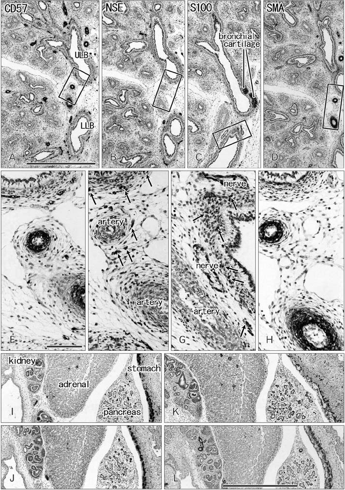
Fig. 1 Immunohistochemistry of the lung, kidney, adrenal, pancreas, and stomach in sections from a specimen at 10 weeks (50-mm crown-rump length). Sagittal sections. Panels A–H show immunostaining of the left lung (A and E, CD57; B and F, neuron-specific enolase [NSE]; C and G, S100; D and H, smooth muscle actin [SMA]), while panels I–L show that of the kidney, adrenal, pancreas and stomach. Panels E–H are higher-magnification views of the squares in panels A–D, re spec tively. Arrows in panels F and G indicate positive nerve elements. Panels I (CD57), J (NSE), K (S100), or L (SMA) displays the same section as shown in panel A, B, C, or D. Panels A–D, panels E–H, and panels I–L were prepared at the same magnification, respec tively. ULB, upper lobar bronchus; LLB, lower lobar bronchus. Scale bars=1 mm (A, L), 0.1 mm (E).
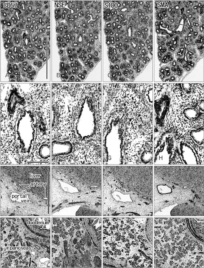
Fig. 2 Immunohistochemistry of the lung, liver, pancreas, and duodenum in sections from a specimen at 15 weeks (110 mm crown-rump length). Sagi ttal sections. Panels A–H show immu nostaining of the right lung lower lobe (A and E, CD57; B and F, neuron-specific enolase [NSE]; C and G, S100; D and H, smooth muscle actin [SMA]). Panels E–H are higher-magnification views of the squares in panels A–D, re spectively. Arrows in panels F and G indicate positive nerve elements. Panels I–L show that of the hilar portion of the liver including hepatic arteries and portal vein. Panels M–O show that of the pancreas and duodenum. Panels I and M (CD57), panels J and N (NSE), panels K and O (S100), or panels L and P (SMA) display the same section as shown in panel A, B, C or D. Panels A–D, panels E–H, and panels I–P were prepared at the same magnification, respectively. Scale bars=1 mm (A, I), 0.1 mm (E).

Fig. 3 Immunohistochemistry of the lung and heart in sections from a specimen at 16 weeks (125 mm crown-rump length). Horizontal sections. Panels A–H show immunostaining of the left lung lower lobe (A and E, CD57; B and F, S100; C and G, neuron-specific enolase [NSE]; D and H, smooth muscle actin [SMA]), while panels I–L show that of the left ventricular wall. Panels E–H are higher-magnification views of the squares in panels A–D, respectively. Panels A and B (or C and D) are adjacent sections. Panel I (CD57), panel J (S100), panel K (NSE), or panel L (SMA) displays the same section as shown in panel A, B, C, or D. Panels A–D, panels E–H, and panels I–L were prepared at the same magnification, respectively. Scale bars=1 mm (A, I), 0.1 mm (E).
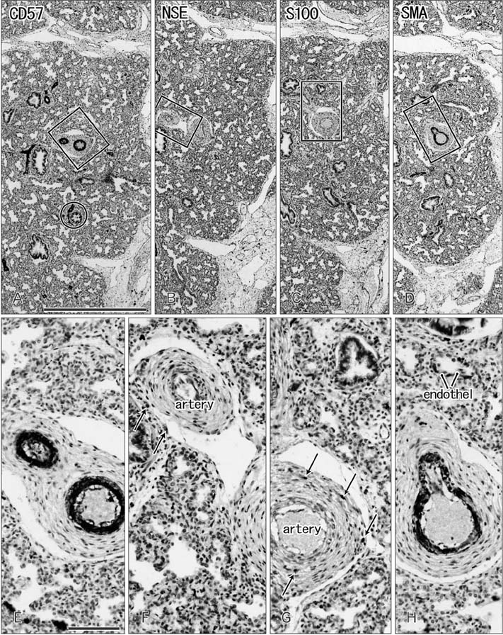
Fig. 4 Immunohistochemistry of the lung in sections from a specimen at 30 weeks (255 mm crown-rump length). Sagittal sections. Panels A–H show immunostaining of the right lung middle lobe (A and E, CD57; B and F, neuron-specific enolase [NSE]; C and G, S100; D and H, smooth muscle actin [SMA]). Panels E–H are higher-magnification views of the squares in panels A–D, respectively. Two circles in panel A are shown in Fig. 5 at higher magnification. Panels A–D and panels E–H were prepared at the same magnification, respectively. Arrows is panel F and G indicate positive nerve elements. Scale bars=1 mm (A), 0.1 mm (E).
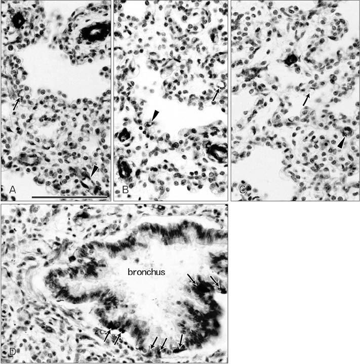
Fig. 5 CD57 reactivity in arterioles and capillaries near and adjacent to alveoli as well as in neuroendocrine cells of the bronchi. Sagittal sections. Panels A–C show peripheral alveolar areas in a specimen of 235 mm crown-rump length (CRL). Panel D displays a higher-magnification view of the circle in Fig. 4A (30 weeks; 255 mm CRL). Panels A–C contain both CD57-positive (arrowheads) and negative (arrows) capillaries. Arterioles are strongly positive. Panel D exhibits a subsegmental bronchus containing CD57-positive neuroendocrine cells (arrows). The other positive structures around the bronchus are supplying nerves: some of them to the bronchus. All panels were prepared at the same magnification. Scale bar=0.1 mm (A).
Reference
-
1. Kleene R, Schachner M. Glycans and neural cell interactions. Nat Rev Neurosci. 2004; 5:195–208.
Article2. Bronner-Fraser M. Analysis of the early stages of trunk neural crest migration in avian embryos using monoclonal antibody HNK-1. Dev Biol. 1986; 115:44–55.
Article3. Verberne ME, Gittenberger-de Groot AC, Poelmann RE. Lineage and development of the parasympathetic nervous system of the embryonic chick heart. Anat Embryol (Berl). 1998; 198:171–184.
Article4. Ikeda T, Iwasaki K, Shimokawa I, Sakai H, Ito H, Matsuo T. Leu-7 immunoreactivity in human and rat embryonic hearts, with special reference to the development of the conduction tissue. Anat Embryol (Berl). 1990; 182:553–562.
Article5. Aoyama N, Tamaki H, Kikawada R, Yamashina S. Development of the conduction system in the rat heart as determined by Leu-7 (HNK-1) immunohistochemistry and computer graphics reconstruction. Lab Invest. 1995; 72:355–366.6. Vassiliadou N, Bulmer JN. Immunohistochemical evidence for increased numbers of ‘classic’ CD57+ natural killer cells in the endometrium of women suffering spontaneous early pregnancy loss. Hum Reprod. 1996; 11:1569–1574.
Article7. O'Rahilly R, Müller F. Human embryology and teratology. 2nd ed. New York: Wiley-Liss;1996. p. 265–271.8. Hayashi S, Murakami G, Ohtsuka A, Itoh M, Nakano T, Fukuzawa Y. Connective tissue configuration in the human liver hilar region with special reference to the liver capsule and vascular sheath. J Hepatobiliary Pancreat Surg. 2008; 15:640–647.
Article9. Miyake N, Hayashi S, Kawase T, Cho BH, Murakami G, Fujimiya M, Kitano H. Fetal anatomy of the human carotid sheath and structures in and around it. Anat Rec (Hoboken). 2010; 293:438–445.
Article10. Haimoto H, Hosoda S, Kato K. Differential distribution of immunoreactive S100-alpha and S100-beta proteins in normal nonnervous human tissues. Lab Invest. 1987; 57:489–498.11. Matsunou H, Konishi F. Papillary-cystic neoplasm of the pancreas: a clinicopathologic study concerning the tumor aging and malignancy of nine cases. Cancer. 1990; 65:283–291.
Article12. Yantiss RK, Chang HK, Farraye FA, Compton CC, Odze RD. Prevalence and prognostic significance of acinar cell differentiation in pancreatic endocrine tumors. Am J Surg Pathol. 2002; 26:893–901.
Article13. Kawamoto A, Kitamura K, Yamamoto M, Murakami G, Abe S, Katori Y. Morphological differences in innervation between mucous glands and serous glands: a quantitative histological study using the sublingual glands of elderly humans. Acta Otolaryngol. 2015; 135:942–949.
Article14. Kaplan RC, Sinclair E, Landay AL, Lurain N, Sharrett AR, Gange SJ, Xue X, Parrinello CM, Hunt P, Deeks SG, Hodis HN. T cell activation predicts carotid artery stiffness among HIV-infected women. Atherosclerosis. 2011; 217:207–213.
Article15. Schumacher H, Kaiser E, Schnabel PA, Sykora J, Eckstein HH, Allenberg JR. Immunophenotypic characterisation of carotid plaque: increased amount of inflammatory cells as an independent predictor for ischaemic symptoms. Eur J Vasc Endovasc Surg. 2001; 21:494–501.
Article16. Winchester R, Giles JT, Nativ S, Downer K, Zhang HZ, Bag-Ozbek A, Zartoshti A, Bokhari S, Bathon JM. Association of elevations of specific T cell and monocyte subpopulations in rheumatoid arthritis with subclinical coronary artery atherosclerosis. Arthritis Rheumatol. 2016; 68:92–102.
Article17. Yu HT, Youn JC, Lee J, Park S, Chi HS, Lee J, Choi C, Park S, Choi D, Ha JW, Shin EC. Characterization of CD8(+)CD57(+) T cells in patients with acute myocardial infarction. Cell Mol Immunol. 2015; 12:466–473.
Article18. Duvar S, Peter-Katalinić J, Hanisch FG, Müthing J. Isolation and structural characterization of glycosphingolipids of in vitro propagated bovine aortic endothelial cells. Glycobiology. 1997; 7:1099–1109.
Article
- Full Text Links
-
- Actions
-
Cited
- CITED
-
- Close
- Share
-
- Similar articles
-
- Oral Administration of Glycine and Leucine Dipeptides Improves Skin Hydration and Elasticity in UVB-Irradiated Hairless Mice
- The Prognostic Significance of Intratumoral Natural Killer Cells in Colorectal Cancer
- Peripheral T Cell Subpopulation in Patients with Pulmonary Tuberculosis
- Association of CD57+ Natural Killer Cells with Better Overall Survival in DLBCL Patients
- Histochemical Studies of Fetal Arteries of Koreans with Special Reference to Atherogenesis in Adults
