Usefulness of shear wave elastography in the diagnosis of oral and maxillofacial diseases
- Affiliations
-
- 1Department of Oral and Maxillofacial Radiology, The Nippon Dental University School of Life Dentistry at Niigata, Niigata, Japan. ogura@ngt.ndu.ac.jp
- 2Advanced Research Center, The Nippon Dental University School of Life Dentistry at Niigata, Niigata, Japan.
- 3Radiology, The Nippon Dental University Niigata Hospital, Niigata, Japan.
- KMID: 2420543
- DOI: http://doi.org/10.5624/isd.2018.48.3.161
Abstract
- PURPOSE
To evaluate the usefulness of shear wave elastography in the diagnosis of oral and maxillofacial diseases.
MATERIALS AND METHODS
Ten patients with oral and maxillofacial diseases and 28 volunteers drawn from our student doctors were examined by shear wave elastography with a 14-MHz linear transducer using an Aplio 300 apparatus (Canon Medical Systems, Otawara, Japan). A statistical analysis of the shear elastic modulus (kPa) of healthy tissue (the sublingual gland, submandibular gland, anterior belly of the digastric muscle, and geniohyoid muscle) in the 28 volunteers was performed using 1-way repeated measures analysis of variance with the Tukey honest significant difference test. The maximum shear elastic modulus (kPa) of 8 patients with squamous cell carcinoma (SCC) and 2 patients with benign lesions was evaluated with the Mann-Whitney U test. The analysis used a 5% significance level.
RESULTS
The mean shear elastic modulus of the sublingual gland (9.4±3.7 kPa) was lower than that of the geniohyoid muscle (19.2±9.2 kPa, P=.000) and the anterior belly of the digastric muscle (15.3±6.1 kPa, P=.004). The maximum shear elastic modulus of the SCCs (109.6±14.4 kPa) was higher than that of the benign lesions (46.4±26.8 kPa, P=.044).
CONCLUSION
Our results demonstrated the usefulness of shear wave elastography in the diagnosis of oral and maxillofacial diseases. Shear wave elastography has the potential to be an effective technique for the objective and quantitative diagnosis of oral and maxillofacial diseases.
MeSH Terms
Figure
-
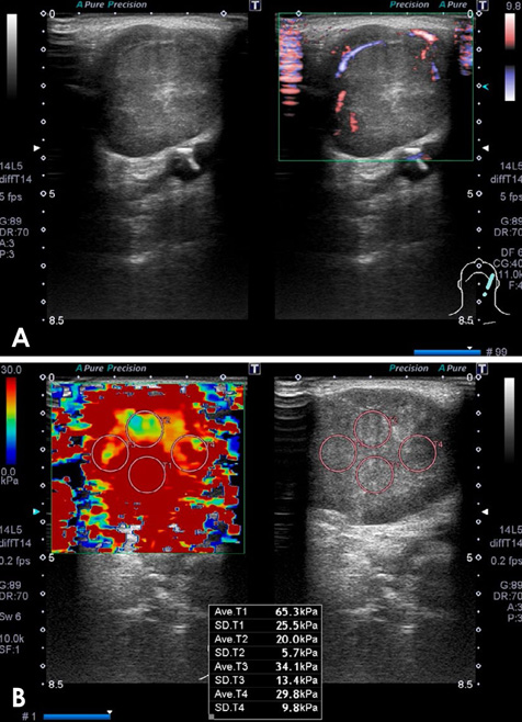
Fig. 1 Ultrasonograms of a pleomorphic adenoma of the left submandibular gland in an 84-year-old man. A. The lesion shows clear boundaries and is isoechoic and heterogeneous, with vascular signals within the lesion. B. T1 (65.3 kPa), T2 (20.0 kPa), T3 (34.1 kPa), and T4 (29.8 kPa) show the shear elastic modulus values of this submandibular gland tumor.
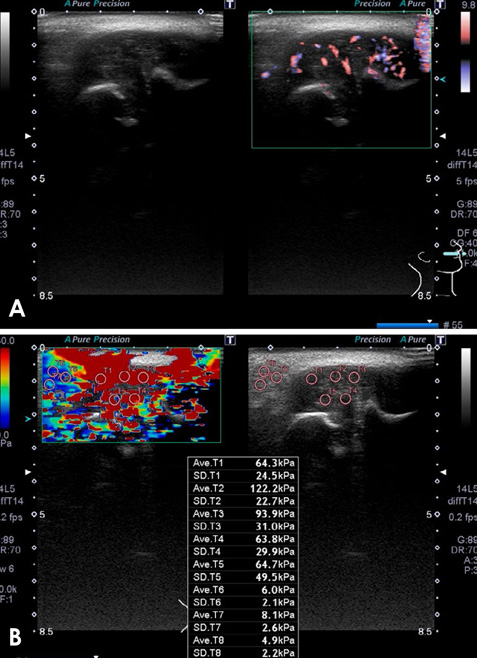
Fig. 2 Ultrasonograms of a squamous cell carcinoma on the right side of the buccal mucosa in a 56-year-old man. A. The lesion shows unclear boundaries, and is isoechoic and heterogeneous, with vascular signals within the lesion. B. T1 (64.3 kPa), T2 (122.2 kPa), T3 (93.9 kPa), and T4 (63.8 kPa) show the shear elastic modulus values of this buccal mucosal tumor.
Cited by 1 articles
-
Strain elastography of palatal tumors in conjunction with intraoral ultrasonography, computed tomography, and magnetic resonance imaging: 2 case reports
Ichiro Ogura, Hiroo Toshima, Tohru Akashiba, Junya Ono, Yasuo Okada
Imaging Sci Dent. 2020;50(1):73-79. doi: 10.5624/isd.2020.50.1.73.
Reference
-
1. Ogura I, Kaneda T, Sasaki Y, Sekiya K, Tokunaga S. Characteristic power Doppler sonographic images of tumorous and non-tumorous buccal space lesions. Dentomaxillofac Radiol. 2013; 42:20120460.
Article2. Acu L, Oktar SÖ, Acu R, Yücel C, Cebeci S. Value of ultrasound elastography in the differential diagnosis of cervical lymph nodes: a comparative study with B-mode and color Doppler sonography. J Ultrasound Med. 2016; 35:2491–2499.3. Turgut E, Celenk C, Tanrivermis Sayit A, Bekci T, Gunbey HP, Aslan K. Efficiency of B-mode ultrasound and strain elastography in differentiating between benign and malignant cervical lymph nodes. Ultrasound Q. 2017; 33:201–207.
Article4. Shingaki M, Nikkuni Y, Katsura K, Ikeda N, Maruyama S, Takagi R, et al. Clinical significance of intraoral strain elastography for diagnosing early stage tongue carcinoma: a preliminary study. Oral Radiol. 2017; 33:204–211.
Article5. Ogura I, Sasaki Y, Sue M, Oda T. Strain elastography of tongue carcinoma using intraoral ultrasonography: a preliminary study to characterize normal tissues and lesions. Imaging Sci Dent. 2018; 48:45–49.
Article6. Bhatia KS, Cho CC, Tong CS, Lee YY, Yuen EH, Ahuja AT. Shear wave elastography of focal salivary gland lesions: preliminary experience in a routine head and neck US clinic. Eur Radiol. 2012; 22:957–965.
Article7. Bhatia KS, Tong CS, Cho CC, Yuen EH, Lee YY, Ahuja AT. Shear wave elastography of thyroid nodules in routine clinical practice: preliminary observations and utility for detecting malignancy. Eur Radiol. 2012; 22:2397–2406.
Article8. Dieterich AV, Andrade RJ, Le Sant G, Falla D, Petzke F, Hug F, et al. Shear wave elastography reveals different degrees of passive and active stiffness of the neck extensor muscles. Eur J Appl Physiol. 2017; 117:171–178.
Article9. Arda K, Ciledag N, Aktas E, Aribas BK, Köse K. Quantitative assessment of normal soft-tissue elasticity using shear-wave ultrasound elastography. AJR Am J Roentgenol. 2011; 197:532–536.
Article10. Azizi G, Keller JM, Mayo ML, Piper K, Puett D, Earp KM, et al. Thyroid nodules and shear wave elastography: a new tool in thyroid cancer detection. Ultrasound Med Biol. 2015; 41:2855–2865.
Article11. Azizi G, Keller JM, Mayo ML, Piper K, Puett D, Earp KM, et al. Shear wave elastography and cervical lymph nodes: predicting malignancy. Ultrasound Med Biol. 2016; 42:1273–1281.
Article12. Cheng KL, Choi YJ, Shim WH, Lee JH, Baek JH. Virtual touch tissue imaging quantification shear wave elastography: prospective assessment of cervical lymph nodes. Ultrasound Med Biol. 2016; 42:378–386.
Article13. Desmots F, Fakhry N, Mancini J, Reyre A, Vidal V, Jacquier A, et al. Shear wave elastography in head and neck lymph node assessment: image quality and diagnostic impact compared with B-mode and Doppler ultrasonography. Ultrasound Med Biol. 2016; 42:387–398.
Article14. Suh CH, Choi YJ, Baek JH, Lee JH. The diagnostic performance of shear wave elastography for malignant cervical lymph nodes: a systematic review and meta-analysis. Eur Radiol. 2017; 27:222–230.
Article15. Ogura I, Amagasa T, Fujii E, Yoshimasu H. Quantitative evaluation of consistency of normal mucosa, leukoplakia and squamous cell carcinoma of the tongue. J Craniomaxillofac Surg. 1998; 26:107–111.
Article
- Full Text Links
-
- Actions
-
Cited
- CITED
-
- Close
- Share
-
- Similar articles
-
- Diagnostic Performance of Quantitative Shear Wave Ultrasound Elastography for Thyroid Cancer
- Ultrasound elastography of the thyroid: principles and current status
- Future of breast elastography
- Shear-Wave Elastography of Segmental Infarction of the Testis
- Shear wave elastography: a systematic review and meta-analysis
