Primary Colonic Follicular Lymphoma Presenting as Four Diminutive Sessile Polyps Found Incidentally During Colonoscopy
- Affiliations
-
- 1Department of Internal Medicine, School of Medicine, Kyungpook National University, Kyungpook National University Hospital, Daegu, Korea. mdleehs@knu.ac.kr
- 2Department of Pathology, School of Medicine, Kyungpook National University, Kyungpook National University Hospital, Daegu, Korea.
- KMID: 2419713
- DOI: http://doi.org/10.5946/ce.2017.114
Abstract
- Follicular lymphomas, which typically arise in the lymph nodes with spleen, liver, and bone marrow involvement, have generally low occurrence rates in Asian countries as compared with Western countries. Follicular lymphomas of the gastrointestinal tract are rare, and primary colonic follicular lymphomas are particularly rare compared with others found in the small intestine and duodenum. Colonoscopic imaging of colonic lymphomas, including follicular lymphoma, may reveal mucosal ulcerations, erosions, indurations, polypoid mass-like lesions, and diffuse mucosal nodularity. Herein, we report a unique case of a follicular lymphoma of the transverse colon characterized by four sessile diminutive polyps located intermittently with multiple lymph node involvement in a 62-year-old man.
Keyword
MeSH Terms
Figure
-

Fig. 1. Colonoscopic findings. (A, B) Sessile polyps in the hepatic flexure and proximal transverse colon were both 2 mm in size. They were removed using forceps. (C-F) Sessile polyps in the proximal and distal transverse colon were 4 and 3 mm in size, respectively. They were resected via endoscopic mucosal resection after injecting saline into the submucosa.
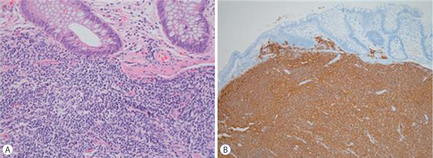
Fig. 2. Histologic examinations of colonic polyps. (A) Homogeneous small lymphoid cells with irregular nuclei are noted (hematoxylin and eosin staining, ×200). (B) Lymphoid cells are diffusely positive for CD20 (immunohistochemical staining for CD20, ×100).

Fig. 3. Positron emission tomography-computed tomography findings. Lymphoma involvement in the spleen and lymph nodes on both sides of the diaphragm is observed.
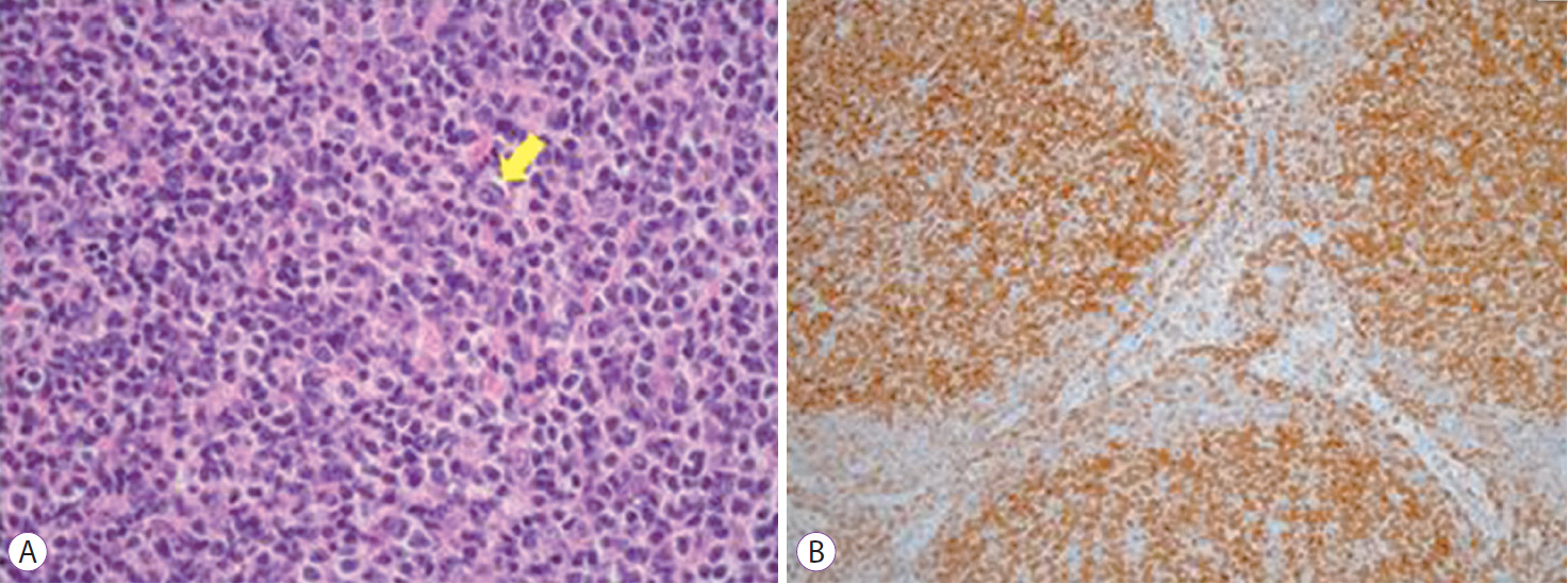
Fig. 4. Histologic examination of the lymph nodes in the left side of the neck. (A) Homogeneous small lymphoid cells with irregular nuclei resembling centrocytes are noted in the follicles. A centroblast is observed (arrow) (hematoxylin and eosin staining, ×400). (B) Lymphoid cells in the follicles are uniformly positive for Bcl-2 (Immunohistochemical staining for Bcl-2, ×100).
Reference
-
1. Zucca E, Roggero E, Bertoni F, Cavalli F. Primary extranodal non-Hodgkin’s lymphomas. Part 1: gastrointestinal, cutaneous and genitourinary lymphomas. Ann Oncol. 1997; 8:727–737.
Article2. Damaj G, Verkarre V, Delmer A, et al. Primary follicular lymphoma of the gastrointestinal tract: a study of 25 cases and a literature review. Ann Oncol. 2003; 14:623–629.
Article3. Iwamuro M, Okada H, Takata K, et al. Colorectal manifestation of follicular lymphoma. Intern Med. 2016; 55:1–8.
Article4. Misdraji J, Harris NL, Hasserjian RP, Lauwers GY, Ferry JA. Primary follicular lymphoma of the gastrointestinal tract. Am J Surg Pathol. 2011; 35:1255–1263.
Article5. Kim YH, Lee JH, Yang SK, et al. Primary colon lymphoma in Korea: a KASID (Korean association for the study of intestinal diseases) study. Dig Dis Sci. 2005; 50:2243–2247.
Article6. Hayashi N, Tanaka S, Hewett DG, et al. Endoscopic prediction of deep submucosal invasive carcinoma: validation of the narrow-band imaging international colorectal endoscopic (NICE) classification. Gastrointest Endosc. 2013; 78:625–632.
Article7. Carbone PP, Kaplan HS, Musshoff K, Smithers DW, Tubiana M. Report of the committee on Hodgkin’s disease staging classification. Cancer Res. 1971; 31:1860–1861.8. Swerdlow SH, Campo E, Harris NL, et al. WHO classification of tumours of haematopoietic and lymphoid tissues. 4th ed. Lyon: International Agency for Research on Cancer;2008.9. Shia J, Teruya-Feldstein J, Pan D, et al. Primary follicular lymphoma of the gastrointestinal tract: a clinical and pathologic study of 26 cases. Am J Surg Pathol. 2002; 26:216–224.10. Ferreira A, Gonçalves R, Rolanda C, et al. A different kind of colon polyp. Gastroenterology. 2012; 143:1440. 1693-1694.
Article11. Takata K, Okada H, Ohmiya N, et al. Primary gastrointestinal follicular lymphoma involving the duodenal second portion is a distinct entity: a multicenter, retrospective analysis in Japan. Cancer Sci. 2011; 102:1532–1536.
Article12. Pyeon SI, Song GA, Baek DH, et al. Primary follicular lymphoma in the rectum incidentally found on screening colonoscopy. Korean J Gastroenterol. 2017; 69:139–142.
Article13. Kwon BS, Kim CD, Park JY, et al. A case of primary follicular lymphoma arising in the rectum. Korean J Gastrointest Endosc. 2006; 33:285–288.14. Wang MH, Wong JM, Lien HC, Lin CW, Wang CY. Colonoscopic manifestations of primary colorectal lymphoma. Endoscopy. 2001; 33:605–609.
Article15. Myung SJ, Joo KR, Yang SK, et al. Clinicopathologic features of ileocolonic malignant lymphoma: analysis according to colonoscopic classification. Gastrointest Endosc. 2003; 57:343–347.
Article16. Kahl BS, Yang DT. Follicular lymphoma: evolving therapeutic strategies. Blood. 2016; 127:2055–2063.
Article17. Salles G, Seymour JF, Offner F, et al. Rituximab maintenance for 2 years in patients with high tumour burden follicular lymphoma responding to rituximab plus chemotherapy (PRIMA): a phase 3, randomised controlled trial. Lancet. 2011; 377:42–51.
Article18. Hassan C, Repici A, Zullo A, Kanakadandi V, Sharma P. Colonic polyps: are we ready to resect and discard? Gastrointest Endosc Clin N Am. 2013; 23:663–678.19. Radaelli F. The resect-and-discard strategy for management of small and diminutive colonic polyps. Gastroenterol Hepatol (N Y). 2013; 9:305–308.20. Hassan C, Pickhardt PJ, Kim DH, et al. Systematic review: distribution of advanced neoplasia according to polyp size at screening colonoscopy. Aliment Pharmacol Ther. 2010; 31:210–217.
Article
- Full Text Links
-
- Actions
-
Cited
- CITED
-
- Close
- Share
-
- Similar articles
-
- Resection of Diminutive and Small Colorectal Polyps: What Is the Optimal Technique?
- Do Distal Colonic Polyps Predict Proximal Adenomas?
- Diminutive Colonic Polyps (Less than 5 mm in diameter) : Endoscopic and Histologic Study
- Primary Follicular Lymphoma in the Rectum Incidentally Found on Screening Colonoscopy
- Clinicopathologic Features and Clinical Significance of Small and Diminutive Colorectal Polyps
