Classical oral manifestations of Dyke-Davidoff-Masson syndrome: a case report with review of the literature
- Affiliations
-
- 1Department of Pediatric & Preventive Dentistry, Government Dental College & Hospital, Nagpur, India. riteshpedo@gmail.com
- 2Department of Oral Medicine & Radiology, Government Dental College & Hospital, Nagpur, India.
- KMID: 2419236
- DOI: http://doi.org/10.5125/jkaoms.2018.44.4.198
Abstract
- Dyke-Davidoff-Masson syndrome is a non-inherited rare condition that presents during childhood and is characterized by seizures, hemiplegia, mental retardation, cerebral hemiatrophy, calvarial thickening, and hyperpneumatization of the frontal sinuses. The present article highlights a case of a 12-year-old male child with additional clinical findings of café-au-late pigmentation and ocular lipodermoid. This is the first case report of Dyke-Davidoff-Masson syndrome to describe oral manifestations, such as unilateral delayed eruption of teeth, hypoplasia, and taurodontism, which could be unique and characteristic of this condition. Oral health care providers and physicians should be aware of these oral observations as dental referrals could warrant early dental prophylactic care and can be useful in diagnosing the possible time of injury and type of Dyke-Davidoff-Masson syndrome.
MeSH Terms
Figure
-
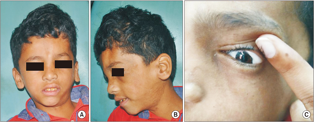
Fig. 1 Extraoral photograph showing facial asymmetry (A), café-au-late pigmentation (B), and lipodermoid of the left eye (C).

Fig. 2 Intraoral photograph showing hypoplastic anterior teeth, severe malocclusion with an open bite (A), and a bucally erupting #35 (B).
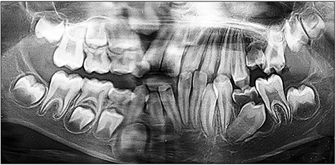
Fig. 3 Panoramic radiograph showing delayed eruption of the right side permanent teeth and taurodontism.
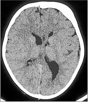
Fig. 4 Computed tomography image of the brain showing mild atrophy of the left cerebral hemisphere, dilatation of the left lateral ventricle, sulcogyral space prominence, and calvarial thickening of the frontal and parietal bones.
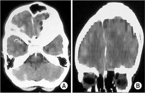
Fig. 5 Computed tomography image of the brain showing left frontal sinus enlargement, thickening of the squamous part of the temporal bone (A), and mild elevation of the left orbital roof (B).
Reference
-
1. Dyke CG, Davidoff LM, Masson CB. Cerebral hemiatrophy and homolateral hypertrophy of the skull and sinuses. Surg Gynecol Obstet. 1933; 57:588–600.2. Aguiar PH, Liu CW, Leitão H, Issa F, Lepski G, Figueiredo EG, et al. MR and CT imaging in the Dyke-Davidoff-Masson syndrome. Report of three cases and contribution to pathogenesis and differential diagnosis. Arq Neuropsiquiatr. 1998; 56:803–807.
Article3. Singh P, Saggar K, Ahluwalia A. Dyke-Davidoff-Masson syndrome: classical imaging findings. J Pediatr Neurosci. 2010; 5:124–125.
Article4. Sener RN, Jinkins JR. MR of craniocerebral hemiatrophy. Clin Imaging. 1992; 16:93–97.
Article5. Stred SE, Byrum CJ, Bove EL, Oliphant M. Coarctation of midaortic arch presenting with monoparesis. Ann Thorac Surg. 1986; 42:210–212.6. Demir Y, Sürücü E, Çilingir V, Bulut MD, Tombul T. Dyke-Davidoff-Masson syndrome with cerebral hypometabolism and unique crossed cerebellar diaschisis in 18F-FDG PET/CT. Clin Nucl Med. 2015; 40:757–758.
Article7. Shrestha B. Acquired cerebral hemiatrophy: Dyke-Davidoff-Masson syndrome--a case report. Turk Neurosurg. 2013; 23:117–121.8. Solomon GE, Hilal SK, Gold AP, Carter S. Natural history of acute hemiplegia of childhood. Brain. 1970; 93:107–120.
Article9. Uduma FU, Emejulu JK, Motah M, Okere PC, Ongolo PC, Muna W. Differential diagnoses of cerebral hemiatrophy in childhood: a review of literature with illustrative report of two cases. Glob J Health Sci. 2013; 5:195–207.
Article10. Arora R, Rani JY. Dyke-Davidoff-Masson syndrome: imaging features with illustration of two cases. Quant Imaging Med Surg. 2015; 5:469–471.11. Dutta A, Bose S, Sen K, Pandit N, Sharma S. Refractory seizure in childhood: Dyke-Davidoff-Masson syndrome revisited. Oman Med J. 2016; 31:304–308.
Article12. Kumar NV, Gugapriya TS, Guru AT, Kumari SN. Dyke-Davidoff-Masson syndrome. Int J Appl Basic Med Res. 2016; 6:57–59.
Article13. Jain D, Aggarwal HK, Goyal S, Mittal A. Dyke-Davidoff-Masson syndrome: a rare case report. Iran J Neurol. 2014; 13:255–256.14. Shen WC, Chen CC, Lee SK, Ho YJ, Lee KR. Magnetic resonance imaging of cerebral hemiatrophy. J Formos Med Assoc. 1993; 92:995–1000.15. Roy U, Panwar A, Mukherjee A, Biswas D. Adult presentation of Dyke-Davidoff-Masson syndrome: a case report. Case Rep Neurol. 2016; 8:20–26.
Article16. Behera MR, Patnaik S, Mohanty AK. Dyke-Davidoff-Masson syndrome. J Neurosci Rural Pract. 2012; 3:411–413.
Article17. Kumar LSV. Café au lait spot: case report. J Adv Clin Res Insights. 2014; 1:106–107.18. Bhallil S, Benatiya I, El Abdouni O, Mahjoubi B, Hicham T. Goldenhar syndrome: ocular features. Bull Soc Belge Ophtalmol. 2010; 17–19.19. Sethi NK, Sethi PK, Torgovnick J, Arsura E. Acquired Dyke-Davidoff-Masson syndrome: a clinicoradiographic correlation. Eastern J Med. 2011; 16:269–271.20. Witkop CJ Jr, Keenan KM, Cervenka J, Jaspers MT. Taurodontism: an anomaly of teeth reflecting disruptive developmental homeostasis. Am J Med Genet Suppl. 1988; 4:85–97.
Article21. Varghese B, Aneesh M, Singh N, Gilwaz P. A case of rasmussen encephalitis: the differential diagnoses and role of diagnostic imaging. Oman Med J. 2014; 29:67–70.
Article22. Auvin S, Bellavoine V, Merdariu D, Delanoë C, Elmaleh-Bergés M, Gressens P, et al. Hemiconvulsion-hemiplegia-epilepsy syndrome: current understandings. Eur J Paediatr Neurol. 2012; 16:413–421.
Article23. Qiu BP, Shi CH. Silver-Russel syndrome: a case report. World J Pediatr. 2007; 3:68–70.24. Moon WK, Chang KH, Kim IO, Han MH, Choi CG, Suh DC, et al. Germinomas of the basal ganglia and thalamus: MR findings and a comparison between MR and CT. AJR Am J Roentgenol. 1994; 162:1413–1417.
Article25. Amor DJ, Kornberg AJ, Smith LJ. Encephalocraniocutaneous lipomatosis (Fishman syndrome): a rare neurocutaneous syndrome. J Paediatr Child Health. 2000; 36:603–605.
Article26. Jacoby CG, Go RT, Hahn FJ. Computed tomography in cerebral hemiatrophy. AJR Am J Roentgenol. 1977; 129:5–9.
Article
- Full Text Links
-
- Actions
-
Cited
- CITED
-
- Close
- Share
-
- Similar articles
-
- A Case of Dyke-Davidoff-Masson Syndrome with Infantile Spasm
- Two cases of Dyke-Davidoff-Masson syndrome
- Craniofacial Deformity in a Patient with Dyke-Davidoff-Masson Syndrome: A Case Report
- Incidentally Detected Acquired Cerebral Hemiatrophy in Old Age: Dyke-Davidoff-Masson Syndrome
- A case of dyke-davidoff-masson syndrome associated with hypopituitarism and diabetes mellitus
