Mismatch between TOF MR Angiography and CT Angiography of the Middle Cerebral Artery may be a Critical Sign in Cerebrovascular Dynamics
- Affiliations
-
- 1Department of Neurosurgery, Washokai Sadamoto Hospital, Ehime, Japan.
- 2Department of Geriatric Medicine and Neurology, Ehime University, Ehime, Japan. migase@m.ehime-u.ac.jp
- KMID: 2418852
- DOI: http://doi.org/10.3349/ymj.2018.59.1.80
Abstract
- PURPOSE
Although time-of-flight (TOF)-magnetic resonance angiography (MRA) can clearly depict intracranial arteries, the arterial flow of middle cerebral artery (MCA) is occasionally not detected. We evaluated this phenomenon with reference to cerebrovascular dynamics.
MATERIALS AND METHODS
Seventeen patients with suspected occlusion of MCA or internal carotid artery on TOF-MRA were enrolled. All patients underwent CT angiography (CTA) and quantitative cerebral blood flow (CBF) examination for measurement of resting CBF and cerebrovascular reactivity (CVR). Depending on appearance, patients were categorized into three groups. Group A (n=6) had MCA delineation on both MRA and CTA, while groups B (n=6) and C (n=5) had no signal on MRA, but Group B had a MCA delineation on CTA.
RESULTS
No significant difference between resting CBF and CBF after the administration of acetazolamide was seen among 3 groups. In contrast, mean CVR in group B was -19.7±18.1%, which was significantly lower than group A [36.4±21.7% (p < 0.05)], but not than group C (21.4±35.2%). Furthermore, all patients in group B displayed a so-called steal phenomenon.
CONCLUSION
This study is the first to show that visualization of MCA on TOF-MRA closely correlates with CVR, and that a vascular pattern showing no MCA signal intensity on MRA but with MCA delineation on CTA indicates a critical cerebrovascular condition.
Keyword
MeSH Terms
Figure
-
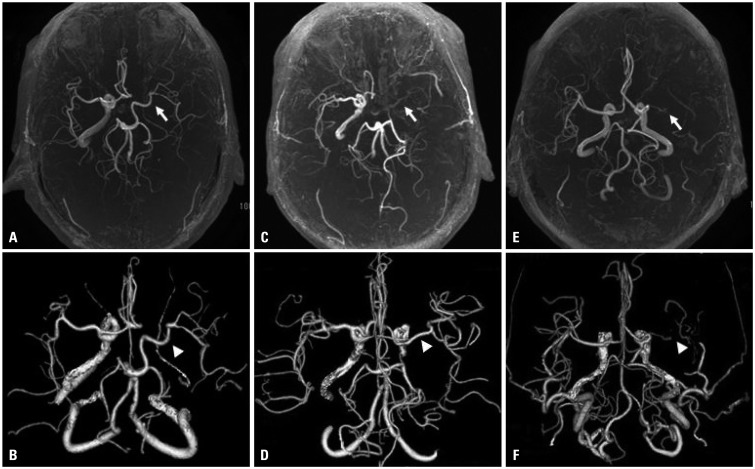
Fig. 1 Categorized 3 groups corresponding to their visualization on TOF-MRA (A, C, and E) and CTA (B, D, and F) are shown. Group A (A and B) had a MCA signal on TOF-MRA (arrow) and an anterograde MCA delineation on CTA (arrowhead); group B (C and D) had no MCA signal (arrow) and MCA delineation (arrowhead); and group C (E and F) had no MCA signal (arrow) and no MCA delineation (arrowhead) on both modalities. TOF-MRA, time-of- flight-magnetic resonance angiography; CTA, CT angiography, MCA, middle cerebral artery.
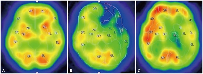
Fig. 2 CBF maps after acetazolamide administration in each group are depicted, coinciding with those of Fig. 1, where group A revealing almost sufficient vascular response in the lesion side, whereas group C showing partly decreased in comparison with contralateral side. Note that CBF maps of group B revealed intensely decreased vascular response in comparison with other 2 groups. CBF, cerebral blood flow.
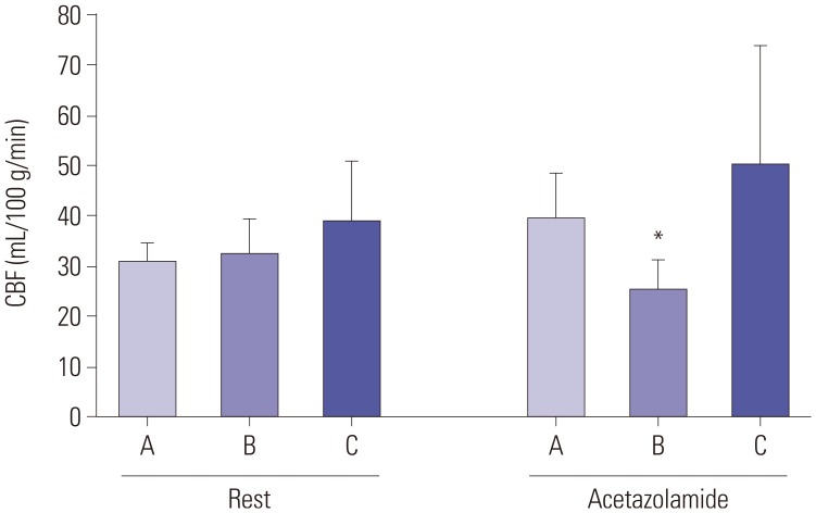
Fig. 3 Mean CBF values of 3 categorized groups (A, B, and C) at rest and after acetazolamide administration were revealed in left and right, respectively. There were no significant differences in both rest and acetazolamide challenge (left and right) among all groups. In respect to CBF values after acetazolamide challenge (right), only group B had significant difference compared with the value before acetazolamide administration (*p<0.05). CBF, cerebral blood flow.
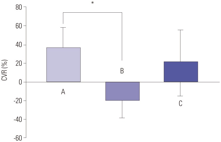
Fig. 4 Mean CVRs in all categorized groups (A, B, and C) are shown. Mean CVR in group B was significantly lower than that in group A (*p<0.05); note that only the mean CVR in group B showed a negative value, which is regarded as a steal phenomenon. CVR: cerebrovascular response.
Reference
-
1. Igase K, Matsubara I, Igase M, Miyazaki H, Sadamoto K. Initial experience in evaluating the prevalence of unruptured intracranial aneurysms detected on 3-tesla MRI. Cerebrovasc Dis. 2012; 33:348–353. PMID: 22378479.
Article2. Choi CG, Lee DH, Lee JH, Pyun HW, Kang DW, Kwon SU, et al. Detection of intracranial atherosclerotic steno-occlusive disease with 3D time-of-flight magnetic resonance angiography with sensitivity encoding at 3T. AJNR Am J Neuroradiol. 2007; 28:439–446. PMID: 17353309.3. Chen H, Li Z, Hong H, Xing S, Liu G, Zhang A, et al. Relationship between visible branch arteries distal to the stenosis on magnetic resonance angiography and stroke recurrence in patients with severe middle cerebral artery trunk stenosis: a one-year follow up study. BMC Neurol. 2015; 15:167. PMID: 26377310.
Article4. Liebeskind DS, Kosinski AS, Lynn MJ, Scalzo F, Fong AK, Fariborz P, et al. Noninvasive fractional flow on MRA predicts stroke risk of intracranial stenosis. J Neuroimaging. 2015; 25:87–91. PMID: 24593693.
Article5. Wilcock DJ, Jaspan T, Worthington BS. Problems and pitfalls of 3-D TOF magnetic resonance angiography of the intracranial circulation. Clin Radiol. 1995; 50:526–532. PMID: 7656518.
Article6. Kim KM, Watabe H, Hayashi T, Hayashida K, Katafuchi T, Enomoto N, et al. Quantitative mapping of basal and vasareactive cerebral blood flow using split-dose 123I-iodoamphetamine and single photon emission computed tomography. Neuroimage. 2006; 33:1126–1135. PMID: 17035048.
Article7. Hirooka R, Ogasawara K, Inoue T, Fujiwara S, Sasaki M, Chida K, et al. Simple assessment of cerebral hemodynamics using single-slab 3D time-of-flight MR angiography in patients with cervical internal carotid artery steno-occlusive diseases: comparison with quantitative perfusion single-photon emission CT. AJNR Am J Neuroradiol. 2009; 30:559–563. PMID: 19039042.
Article8. de Boorder MJ, van der Grond J, van Dongen AJ, Klijn CJ, Jaap Kappelle L, Van Rijk PP, et al. Spect measurements of regional cerebral perfusion and carbondioxide reactivity: correlation with cerebral collaterals in internal carotid artery occlusive disease. J Neurol. 2006; 253:1285–1291. PMID: 17063318.
Article9. Hendrikse J, Klijn CJ, van Huffelen AC, Kappelle LJ, van der Grond J. Diagnosing cerebral collateral flow patterns: accuracy of non-invasive testing. Cerebrovasc Dis. 2008; 25:430–437. PMID: 18349537.
Article10. Kusunoki K, Oka Y, Saito M, Sadamoto K, Sakaki S, Miki H, et al. Changes in visibility of intracranial arteries on MRA with normal ageing. Neuroradiology. 1999; 41:813–819. PMID: 10602853.
Article11. Ishimaru H, Ochi M, Morikawa M, Takahata H, Matsuoka Y, Koshiishi T, et al. Accuracy of pre- and postcontrast 3D time-of-flight MR angiography in patients with acute ischemic stroke: correlation with catheter angiography. AJNR Am J Neuroradiol. 2007; 28:923–926. PMID: 17494671.
- Full Text Links
-
- Actions
-
Cited
- CITED
-
- Close
- Share
-
- Similar articles
-
- Magnetization Transfer Contrast Angiography for Organized Thrombosed Intracranial Aneurysm in TOF MR Angiography: a Case Report
- The Usefulness of 3T-TOF MR angiography in Patients with Cerebral Infarction
- Middle Cerebral Artery Fenestration Associated with an Aneurysm: Case Report
- Infraoptic Anterior Cerebral Artery Arising from Contralateral Internal Carotid Artery: Case Report
- Onyx Embolization of Intracranial Pial Arteriovenous Fistula
