Disruption of the Gut Ecosystem by Antibiotics
- Affiliations
-
- 1Department of Microbiology and Immunology, Brain Korea 21 Project for Medical Sciences, Seoul, Korea. sangsun_yoon@yuhs.ac
- 2Institute for Immunology and Immunological Diseases, Yonsei University College of Medicine, Seoul, Korea.
- KMID: 2418842
- DOI: http://doi.org/10.3349/ymj.2018.59.1.4
Abstract
- The intestinal microbiota is a complex ecosystem consisting of various microorganisms that expands human genetic repertoire and therefore affects human health and disease. The metabolic processes and signal transduction pathways of the host and intestinal microorganisms are intimately linked, and abnormal progression of each process leads to changes in the intestinal environment. Alterations in microbial communities lead to changes in functional structures based on the metabolites produced in the gut, and these environmental changes result in various bacterial infections and chronic enteric inflammatory diseases. Here, we illustrate how antibiotics are associated with an increased risk of antibiotic-associated diseases by driving intestinal environment changes that favor the proliferation and virulence of pathogens. Understanding the pathogenesis caused by antibiotics would be a crucial key to the treatment of antibiotic-associated diseases by mitigating changes in the intestinal environment and restoring it to its original state.
Keyword
MeSH Terms
Figure
-
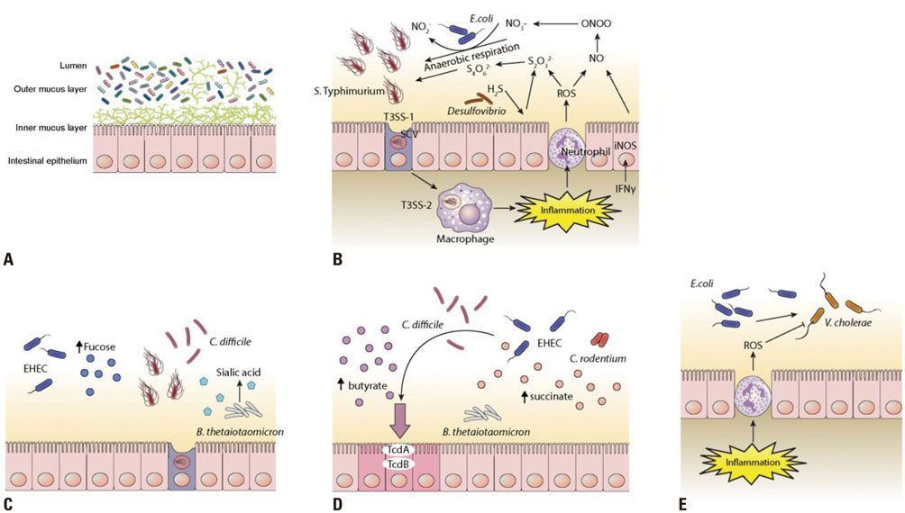
Fig. 1 Pathogens exploit the antibiotic-induced inflammatory conditions. Pathogens use sugars and inorganic compounds generated by the intestinal microbiota as carbon or energy sources and perform anaerobic respiration in the inflammatory conditions caused by antibiotics. (A) When the distribution of intestinal microorganisms is in a stable state, the invasion of pathogenic bacteria is suppressed by antimicrobial substances produced from intestinal bacteria and host cells and the inflammation is suitably controlled. (B) In an inflammatory condition, the colonization of E. coli and Salmonella expands through anaerobic respiration using ROS and RNS, which are released by DUOX2 and iNOS in epithelial cells. Hydrogen sulfide derived from sulfate-reducing bacteria such as Desulfovibrio spp. is converted to thiosulfate during cellular respiration in colonic epithelial cells. ROS generated by neutrophils convert the thiosulfate into tetrathionate that can be used as an electron acceptor. During this process, the generated tetrathionate boosts the growth of S. Typhimurium through tetrathionate respiration that converts tetrathionate to thiosulfate. E. coli reduces nitrate to nitrite through nitrate respiration. (C) Bacteroides thetaiotaomicron decomposes mucosal glycoconjugates to produce sialic acid. EHEC and Salmonella can use the sialic acid as a carbon source. Inflammatory conditions lead to release of fucose from host glycan and the liberated fucose is subsequently consumed by pathogens. As an example, EHEC are known to regulate the expression of virulence genes by sensing the fucose. (D) C. difficile, C. rodentium, and EHEC utilize succinate, which is produced by other intestinal microorganisms. SCFAs excreted during polysaccharide metabolism by aerobic bacteria and butyrate, propionate, and acetate are predominantly present in the intestinal environment. A commensal bacterium, Bacteroides spp., mainly distributes succinate, which is subsequently consumed by secondary fermentative microbes in a steady state and therefore rarely accumulates in the intestinal environment. However, succinate is not consumed under antibiotic treatment or inflammatory conditions, eventually leading to its accumulation in the intestinal lumen. Succinate promotes gluconeogenesis of EHEC. In addition, the colonization and proliferation of C. rodentium are enhanced, especially with expression of virulence genes of the LEE. C. difficile can couple succinate metabolism and convert it to butyrate with the fermentation of carbohydrates, thereby enhancing its colonization and virulence. (E) Antibiotics can trigger the growth of Enterobacteriaceae. ROS at high concentrations result in an expansion of E. coli harboring an extra catalase that are genetically generated through chromosomal modification and eventually favor intestinal colonization of Vibrio cholerae, a strain that is highly sensitive strain to ROS, by reducing the ROS that are excessively generated in inflammatory conditions. E. coli, Escherichia coli; ROS, reactive oxygen species; RNS, reactive nitrogen species; iNOS, inducible nitric oxide synthase; EHEC, Enterohemorrhagic Escherichia coli; C. difficile, Clostridium difficile; C. rodentium, Citrobacter rodentium; SCFAs, short-chain fatty acids; LEE, locus of enterocyte effacement.
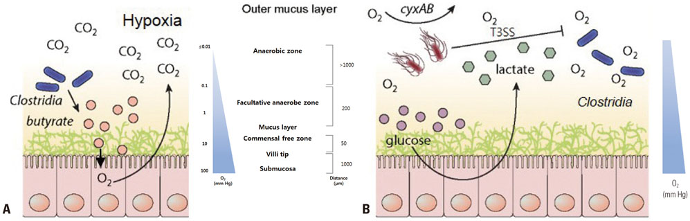
Fig. 2 Effects of antibiotics on the hypoxia barrier of intestinal epithelial cells. (A) In normal conditions, oxygen tension decreases steadily from the intestinal submucosal layer to the lumen. Although the partial pressure of oxygen is approximately 100 mm Hg in the basal layer, it is almost 0 mm Hg in the lumen. Under antibiotic treatment or inflammatory conditions, Clostridia produce butyrate and colonic epithelial cells convert the butyrate to carbon dioxide, leading to maintenance of hypoxia in the lumen. (B) When butyrate is lacking in the intestine, the cells utilize glucose for cellular respiration and the lactate that is released during the process increases oxygenation within the lumen. S. Typhimurium can proliferate using cytochrome bd-II oxidase encoded in cyxB, which is highly expressed at low oxygen concentrations.
Reference
-
1. Kumar H, Lund R, Laiho A, Lundelin K, Ley RE, Isolauri E, et al. Gut microbiota as an epigenetic regulator: pilot study based on whole-genome methylation analysis. MBio. 2014; 5:e02113–e02114.
Article2. Ha CW, Lam YY, Holmes AJ. Mechanistic links between gut microbial community dynamics, microbial functions and metabolic health. World J Gastroenterol. 2014; 20:16498–16517.
Article3. Yang BG, Hur KY, Lee MS. Alterations in gut microbiota and immunity by dietary fat. Yonsei Med J. 2017; 58:1083–1091.
Article4. Sommer F, Anderson JM, Bharti R, Raes J, Rosenstiel P. The resilience of the intestinal microbiota influences health and disease. Nat Rev Microbiol. 2017; 15:630–638.
Article5. Naeem S, Li S. Biodiversity enhances ecosystem reliability. Nature. 1997; 390:507–509.
Article6. Guarner F, Malagelada JR. Gut flora in health and disease. Lancet. 2003; 361:512–519.
Article7. Faith JJ, Guruge JL, Charbonneau M, Subramanian S, Seedorf H, Goodman AL, et al. The long-term stability of the human gut microbiota. Science. 2013; 341:1237439.
Article8. Clemente JC, Ursell LK, Parfrey LW, Knight R. The impact of the gut microbiota on human health: an integrative view. Cell. 2012; 148:1258–1270.
Article9. Kamada N, Seo SU, Chen GY, Núñez G. Role of the gut microbiota in immunity and inflammatory disease. Nat Rev Immunol. 2013; 13:321–335.
Article10. Kamada N, Chen GY, Inohara N, Núñez G. Control of pathogens and pathobionts by the gut microbiota. Nat Immunol. 2013; 14:685–690.
Article11. Turnbaugh PJ, Bäckhed F, Fulton L, Gordon JI. Diet-induced obesity is linked to marked but reversible alterations in the mouse distal gut microbiome. Cell Host Microbe. 2008; 3:213–223.
Article12. Lozupone CA, Stombaugh JI, Gordon JI, Jansson JK, Knight R. Diversity, stability and resilience of the human gut microbiota. Nature. 2012; 489:220–230.
Article13. Tamburini S, Shen N, Wu HC, Clemente JC. The microbiome in early life: implications for health outcomes. Nat Med. 2016; 22:713–722.
Article14. Moya A, Ferrer M. Functional redundancy-induced stability of gut microbiota subjected to disturbance. Trends Microbiol. 2016; 24:402–413.
Article15. Willing BP, Russell SL, Finlay BB. Shifting the balance: antibiotic effects on host-microbiota mutualism. Nat Rev Microbiol. 2011; 9:233–243.
Article16. Sekirov I, Tam NM, Jogova M, Robertson ML, Li Y, Lupp C, et al. Antibiotic-induced perturbations of the intestinal microbiota alter host susceptibility to enteric infection. Infect Immun. 2008; 76:4726–4736.
Article17. Potgieter M, Bester J, Kell DB, Pretorius E. The dormant blood microbiome in chronic, inflammatory diseases. FEMS Microbiol Rev. 2015; 39:567–591.
Article18. Allison SD, Martiny JB. Colloquium paper: resistance, resilience, and redundancy in microbial communities. Proc Natl Acad Sci U S A. 2008; 105:Suppl 1. 11512–11519.19. Becattini S, Taur Y, Pamer EG. Antibiotic-induced changes in the intestinal microbiota and disease. Trends Mol Med. 2016; 22:458–478.
Article20. Pamer EG. Resurrecting the intestinal microbiota to combat antibiotic-resistant pathogens. Science. 2016; 352:535–538.
Article21. Comte J, Fauteux L, Del Giorgio PA. Links between metabolic plasticity and functional redundancy in freshwater bacterioplankton communities. Front Microbiol. 2013; 4:112.
Article22. Modi SR, Collins JJ, Relman DA. Antibiotics and the gut microbiota. J Clin Invest. 2014; 124:4212–4218.
Article23. Langdon A, Crook N, Dantas G. The effects of antibiotics on the microbiome throughout development and alternative approaches for therapeutic modulation. Genome Med. 2016; 8:39.
Article24. DiGiulio DB, Romero R, Amogan HP, Kusanovic JP, Bik EM, Gotsch F, et al. Microbial prevalence, diversity and abundance in amniotic fluid during preterm labor: a molecular and culture-based investigation. PLoS One. 2008; 3:e3056.
Article25. Bohnhoff M, Drake BL, Miller CP. Effect of streptomycin on susceptibility of intestinal tract to experimental Salmonella infection. Proc Soc Exp Biol Med. 1954; 86:132–137.
Article26. Panda S, El khader I, Casellas F, López Vivancos J, García Cors M, Santiago A, et al. Short-term effect of antibiotics on human gut microbiota. PLoS One. 2014; 9:e95476.
Article27. Perez-Lopez A, Behnsen J, Nuccio SP, Raffatellu M. Mucosal immunity to pathogenic intestinal bacteria. Nat Rev Immunol. 2016; 16:135–148.
Article28. Schubert AM, Sinani H, Schloss PD. Antibiotic-induced alterations of the murine gut microbiota and subsequent effects on colonization resistance against Clostridium difficile. MBio. 2015; 6:e00974.
Article29. Prudhomme M, Attaiech L, Sanchez G, Martin B, Claverys JP. Antibiotic stress induces genetic transformability in the human pathogen Streptococcus pneumoniae. Science. 2006; 313:89–92.
Article30. Gipponi M, Sciutto C, Accornero L, Bonassi S, Raso C, Vignolo C, et al. Assessing modifications of the intestinal bacterial flora in patients on long-term oral treatment with bacampicillin or amoxicillin: a random study. Chemioterapia. 1985; 4:214–217.31. Nord CE, Sillerström E, Wahlund E. Effect of tigecycline on normal oropharyngeal and intestinal microflora. Antimicrob Agents Chemother. 2006; 50:3375–3380.
Article32. Jernberg C, Löfmark S, Edlund C, Jansson JK. Long-term ecological impacts of antibiotic administration on the human intestinal microbiota. ISME J. 2007; 1:56–66.
Article33. Jakobsson HE, Jernberg C, Andersson AF, Sjölund-Karlsson M, Jansson JK, Engstrand L. Short-term antibiotic treatment has differing long-term impacts on the human throat and gut microbiome. PLoS One. 2010; 5:e9836.
Article34. Gibson MK, Wang B, Ahmadi S, Burnham CA, Tarr PI, Warner BB, et al. Developmental dynamics of the preterm infant gut microbiota and antibiotic resistome. Nat Microbiol. 2016; 1:16024.
Article35. McFarland LV. Use of probiotics to correct dysbiosis of normal microbiota following disease or disruptive events: a systematic review. BMJ Open. 2014; 4:e005047.
Article36. Dethlefsen L, Huse S, Sogin ML, Relman DA. The pervasive effects of an antibiotic on the human gut microbiota, as revealed by deep 16S rRNA sequencing. PLoS Biol. 2008; 6:e280.
Article37. Dethlefsen L, Relman DA. Incomplete recovery and individualized responses of the human distal gut microbiota to repeated antibiotic perturbation. Proc Natl Acad Sci U S A. 2011; 108:Suppl 1. 4554–4561.
Article38. Hill DA, Hoffmann C, Abt MC, Du Y, Kobuley D, Kirn TJ, et al. Metagenomic analyses reveal antibiotic-induced temporal and spatial changes in intestinal microbiota with associated alterations in immune cell homeostasis. Mucosal Immunol. 2010; 3:148–158.
Article39. Pérez-Cobas AE, Gosalbes MJ, Friedrichs A, Knecht H, Artacho A, Eismann K, et al. Gut microbiota disturbance during antibiotic therapy: a multi-omic approach. Gut. 2013; 62:1591–1601.
Article40. Garrett WS, Gallini CA, Yatsunenko T, Michaud M, DuBois A, Delaney ML, et al. Enterobacteriaceae act in concert with the gut microbiota to induce spontaneous and maternally transmitted colitis. Cell Host Microbe. 2010; 8:292–300.
Article41. Spees AM, Wangdi T, Lopez CA, Kingsbury DD, Xavier MN, Winter SE, et al. Streptomycin-induced inflammation enhances Escherichia coli gut colonization through nitrate respiration. MBio. 2013; 4:e00430–e00413.
Article42. Winter SE, Winter MG, Xavier MN, Thiennimitr P, Poon V, Keestra AM, et al. Host-derived nitrate boosts growth of E. coli in the inflamed gut. Science. 2013; 339:708–711.
Article43. Yoon MY, Min KB, Lee KM, Yoon Y, Kim Y, Oh YT, et al. A single gene of a commensal microbe affects host susceptibility to enteric infection. Nat Commun. 2016; 7:11606.
Article44. Li M, Wang B, Zhang M, Rantalainen M, Wang S, Zhou H, et al. Symbiotic gut microbes modulate human metabolic phenotypes. Proc Natl Acad Sci U S A. 2008; 105:2117–2122.
Article45. Kelly CJ, Zheng L, Campbell EL, Saeedi B, Scholz CC, Bayless AJ, et al. Crosstalk between microbiota-derived short-chain fatty acids and intestinal epithelial HIF augments tissue barrier function. Cell Host Microbe. 2015; 17:662–671.
Article46. Samuel BS, Gordon JI. A humanized gnotobiotic mouse model of host-archaeal-bacterial mutualism. Proc Natl Acad Sci U S A. 2006; 103:10011–10016.
Article47. Belenguer A, Duncan SH, Calder AG, Holtrop G, Louis P, Lobley GE, et al. Two routes of metabolic cross-feeding between Bifidobacterium adolescentis and butyrate-producing anaerobes from the human gut. Appl Environ Microbiol. 2006; 72:3593–3599.
Article48. Ley RE. Harnessing microbiota to kill a pathogen: the sweet tooth of Clostridium difficile. Nat Med. 2014; 20:248–249.
Article49. Ng KM, Ferreyra JA, Higginbottom SK, Lynch JB, Kashyap PC, Gopinath S, et al. Microbiota-liberated host sugars facilitate post-antibiotic expansion of enteric pathogens. Nature. 2013; 502:96–99.
Article50. Ferreyra JA, Wu KJ, Hryckowian AJ, Bouley DM, Weimer BC, Sonnenburg JL. Gut microbiota-produced succinate promotes C. difficile infection after antibiotic treatment or motility disturbance. Cell Host Microbe. 2014; 16:770–777.
Article51. Xu J, Gordon JI. Honor thy symbionts. Proc Natl Acad Sci U S A. 2003; 100:10452–10459.
Article52. Curtis MM, Hu Z, Klimko C, Narayanan S, Deberardinis R, Sperandio V. The gut commensal Bacteroides thetaiotaomicron exacerbates enteric infection through modification of the metabolic landscape. Cell Host Microbe. 2014; 16:759–769.
Article53. Winter SE, Thiennimitr P, Winter MG, Butler BP, Huseby DL, Crawford RW, et al. Gut inflammation provides a respiratory electron acceptor for Salmonella. Nature. 2010; 467:426–429.
Article54. Rivera-Chávez F, Zhang LF, Faber F, Lopez CA, Byndloss MX, Olsan EE, et al. Depletion of butyrate-producing Clostridia from the gut microbiota drives an aerobic luminal expansion of Salmonella. Cell Host Microbe. 2016; 19:443–454.
Article55. Lopez CA, Rivera-Chávez F, Byndloss MX, Bäumler AJ. The periplasmic nitrate reductase NapABC supports luminal growth of Salmonella enterica serovar Typhimurium during colitis. Infect Immun. 2015; 83:3470–3478.
Article56. El Kaoutari A, Armougom F, Gordon JI, Raoult D, Henrissat B. The abundance and variety of carbohydrate-active enzymes in the human gut microbiota. Nat Rev Microbiol. 2013; 11:497–504.
Article57. Donohoe DR, Garge N, Zhang X, Sun W, O'Connell TM, Bunger MK, et al. The microbiome and butyrate regulate energy metabolism and autophagy in the mammalian colon. Cell Metab. 2011; 13:517–526.
Article58. Espey MG. Role of oxygen gradients in shaping redox relationships between the human intestine and its microbiota. Free Radic Biol Med. 2013; 55:130–140.
Article59. Lessa FC, Mu Y, Bamberg WM, Beldavs ZG, Dumyati GK, Dunn JR, et al. Burden of Clostridium difficile infection in the United States. N Engl J Med. 2015; 372:825–834.
Article60. Kelly CP, Pothoulakis C, LaMont JT. Clostridium difficile colitis. N Engl J Med. 1994; 330:257–262.
Article61. Johnston BC, Ma SS, Goldenberg JZ, Thorlund K, Vandvik PO, Loeb M, et al. Probiotics for the prevention of Clostridium difficile-associated diarrhea: a systematic review and meta-analysis. Ann Intern Med. 2012; 157:878–888.
Article62. Martz SL, McDonald JA, Sun J, Zhang YG, Gloor GB, Noordhof C, et al. Administration of defined microbiota is protective in a murine Salmonella infection model. Sci Rep. 2015; 5:16094.
Article63. Bakken JS, Borody T, Brandt LJ, Brill JV, Demarco DC, Franzos MA, et al. Treating Clostridium difficile infection with fecal microbiota transplantation. Clin Gastroenterol Hepatol. 2011; 9:1044–1049.
Article64. Borody TJ, Khoruts A. Fecal microbiota transplantation and emerging applications. Nat Rev Gastroenterol Hepatol. 2011; 9:88–96.
Article65. Hassett DJ, Elkins JG, Ma JF, McDermott TR. Pseudomonas aeruginosa biofilm sensitivity to biocides: use of hydrogen peroxide as model antimicrobial agent for examining resistance mechanisms. Methods Enzymol. 1999; 310:599–608.66. Schultz M. Clinical use of E. coli Nissle 1917 in inflammatory bowel disease. Inflamm Bowel Dis. 2008; 14:1012–1018.
Article67. Hempel S, Newberry SJ, Maher AR, Wang Z, Miles JN, Shanman R, et al. Probiotics for the prevention and treatment of antibiotic-associated diarrhea: a systematic review and meta-analysis. JAMA. 2012; 307:1959–1969.
Article68. Gareau MG, Sherman PM, Walker WA. Probiotics and the gut microbiota in intestinal health and disease. Nat Rev Gastroenterol Hepatol. 2010; 7:503–514.
Article
- Full Text Links
-
- Actions
-
Cited
- CITED
-
- Close
- Share
-
- Similar articles
-
- Microbial Modulation in Inflammatory Bowel Diseases
- Gut-Brain Connection: Microbiome, Gut Barrier, and Environmental Sensors
- Host-microbial Cross-talk in Inflammatory Bowel Disease
- Mental Disorders Linked to Crosstalk between The Gut Microbiome and The Brain
- The clinical impact of gut microbiota in chronic kidney disease
