Complementary Role of Elastography Using Carotid Artery Pulsation in the Ultrasonographic Assessment of Thyroid Nodules: A Prospective Study
- Affiliations
-
- 1Department of Radiology and Center for Imaging Science, Thyroid Center, Samsung Medical Center, Sungkyunkwan University School of Medicine, Seoul 06351, Korea. helena35@hanmail.net
- KMID: 2418563
- DOI: http://doi.org/10.3348/kjr.2018.19.5.992
Abstract
OBJECTIVE
The aim of this study was to evaluate the diagnostic performance of gray-scale ultrasonography (US), Doppler scan, and elastography using carotid artery pulsation in the diagnosis of thyroid nodules and to find a complementary role of elastography.
MATERIALS AND METHODS
A total 197 thyroid nodules with 91 malignant and 106 benign pathologic results from 187 patients (41 males and 146 females; age range, 20-83 years; mean age, 49.4 years) were included in this prospective study. The gray-scale, Doppler US images, elastography with elasticity contrast index (ECI), and stiffness color were assessed. The diagnostic performances of each dataset were assessed in order to differentiate benign from malignant thyroid nodules.
RESULTS
The optimal cut-off value of the ECI was 1.71. The area under receiver operating characteristic curve (Az value) was 0.821 for gray-scale US, 0.661 for the ECI, 0.592 for stiffness color, and 0.539 for Doppler US. The Az value for a combined assessment of gray-scale US and the ECI was higher than that for the gray-scale US alone; however, there was no statistical difference between the two (p = 0.219). The median ECI values of follicular thyroid carcinoma (FTC) and follicular variant of papillary thyroid carcinoma (FVPTC) were significantly lower than those of the other malignant lesions (p = 0.005). Meanwhile, the diffuse sclerosing variant of PTC and a metastatic nodule showed the two highest median values of the ECI.
CONCLUSION
For differentiating thyroid nodules, the diagnostic performances of the combination of gray-scale US and elastography with the ECI were similar to, but not superior, to those of gray-scale US alone. FVPTC and FTC have a significantly lower ECI value than those of the other malignant lesions.
Keyword
MeSH Terms
Figure
-
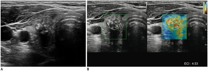
Fig. 1 Gray-scale US (A) and elastography (B) of malignant thyroid nodule.Gray-scale US images (A) show solid hypoechoic nodule with spiculated margins and calcifications in right lobe of thyroid gland. This nodule was classified as K-TIRADS category 5. At elastography evaluation (B), this nodule presents stiff pattern (red) in elastography ROI (green box) and high elasticity contrast index (4.53) calculated from ROI (white octagon). This nodule was confirmed as papillary thyroid carcinoma after surgery. ECI = elasticity contrast index, K-TIRADS = Korean Thyroid Imaging Reporting and Data System, ROI = region of interest, US = ultrasonography
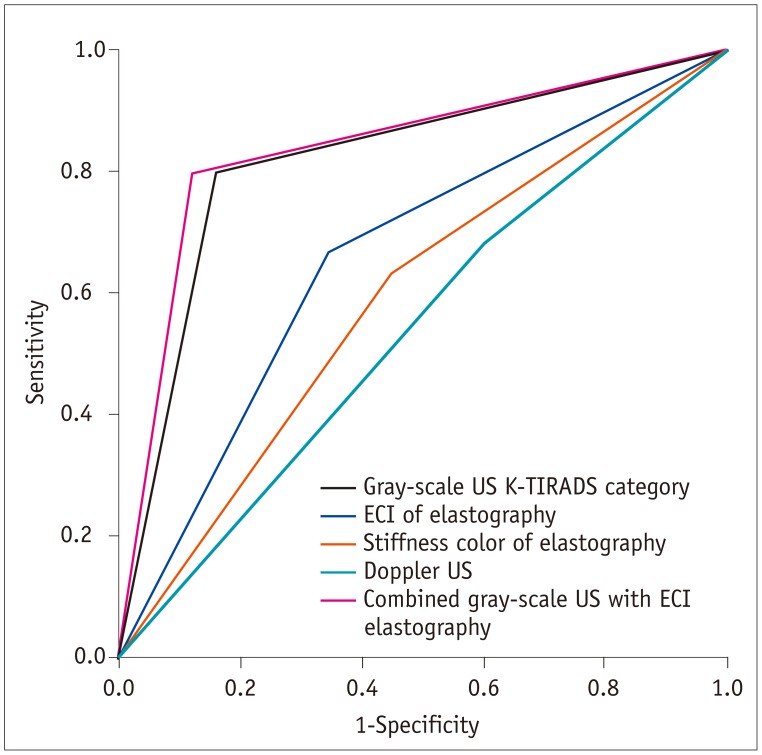
Fig. 2 Receiver operating characteristic curves for gray-scale US K-TIRADS category, ECI and stiffness color of elastography, Doppler US, and combined gray-scale US with ECI elastography for differentiation of benign from malignant thyroid nodules.Az value was 0.821 (95% CI 0.759–0.883) for K-TIRADS category of gray-scale US, 0.661 (95% CI 0.584–0.737) for ECI of elastography, 0.592 (95% CI 0.513–0.672) for stiffness color of elastography, 0.539 (95% CI 0.458–0.619) for Doppler US, and 0.840 (95% CI 0.780–0.900) for combined gray-scale US with ECI elastography, respectively. Az = area under receiver operating characteristics curve, CI = confidence interval
Reference
-
1. Fish SA, Langer JE, Mandel SJ. Sonographic imaging of thyroid nodules and cervical lymph nodes. Endocrinol Metab Clin North Am. 2008; 37:401–417. ixPMID: 18502334.
Article2. Horvath E, Majlis S, Rossi R, Franco C, Niedmann JP, Castro A, et al. An ultrasonogram reporting system for thyroid nodules stratifying cancer risk for clinical management. J Clin Endocrinol Metab. 2009; 94:1748–1751. PMID: 19276237.
Article3. Seo H, Na DG, Kim JH, Kim KW, Yoon JW. Ultrasound-based risk stratification for malignancy in thyroid nodules: a four-Tier Categorization System. Eur Radiol. 2015; 25:2153–2162. PMID: 25680723.
Article4. Park JY, Lee HJ, Jang HW, Kim HK, Yi JH, Lee W, et al. A proposal for a thyroid imaging reporting and data system for ultrasound features of thyroid carcinoma. Thyroid. 2009; 19:1257–1264. PMID: 19754280.
Article5. Kwak JY, Han KH, Yoon JH, Moon HJ, Son EJ, Park SH, et al. Thyroid imaging reporting and data system for US features of nodules: a step in establishing better stratification of cancer risk. Radiology. 2011; 260:892–899. PMID: 21771959.
Article6. Kwak JY, Jung I, Baek JH, Baek SM, Choi N, Choi YJ, et al. Korean Society of Thyroid Radiology (KSThR). Korean Society of Radiology. Image reporting and characterization system for ultrasound features of thyroid nodules: multicentric Korean retrospective study. Korean J Radiol. 2013; 14:110–117. PMID: 23323040.
Article7. Haugen BR, Alexander EK, Bible KC, Doherty GM, Mandel SJ, Nikiforov YE, et al. 2015 American Thyroid Association management guidelines for adult patients with thyroid nodules and differentiated thyroid cancer: the American Thyroid Association guidelines task force on thyroid nodules and differentiated thyroid cancer. Thyroid. 2016; 26:1–133. PMID: 26462967.
Article8. Park JM, Choi Y, Kwag HJ. Partially cystic thyroid nodules: ultrasound findings of malignancy. Korean J Radiol. 2012; 13:530–535. PMID: 22977318.
Article9. National Comprehensive Cancer Network, Inc. 2014 Practice Guidelines in Oncology-Thyroid Carcinoma v.2. Web site. Accessed April 9, 2018. http://www.nccn.org/professionals/physician_gls/f_guidelines.asp.10. Shin JH, Baek JH, Chung J, Ha EJ, Kim JH, Lee YH, et al. Korean Society of Thyroid Radiology (KSThR) and Korean Society of Radiology. Ultrasonography diagnosis and imaging-based management of thyroid nodules: revised Korean Society of Thyroid Radiology consensus statement and recommendations. Korean J Radiol. 2016; 17:370–395. PMID: 27134526.
Article11. Papini E, Guglielmi R, Bianchini A, Crescenzi A, Taccogna S, Nardi F, et al. Risk of malignancy in nonpalpable thyroid nodules: predictive value of ultrasound and color-Doppler features. J Clin Endocrinol Metab. 2002; 87:1941–1946. PMID: 11994321.
Article12. Iared W, Shigueoka DC, Cristófoli JC, Andriolo R, Atallah AN, Ajzen SA, et al. Use of color Doppler ultrasonography for the prediction of malignancy in follicular thyroid neoplasms: systematic review and meta-analysis. J Ultrasound Med. 2010; 29:419–425. PMID: 20194937.13. Gharib H, Papini E, Paschke R, Duick DS, Valcavi R, Hegedüs L, et al. AACE/AME/ETA Task Force on Thyroid Nodules. American Association of Clinical Endocrinologists, Associazione Medici Endocrinologi, and EuropeanThyroid Association medical guidelines for clinical practice for the diagnosis and management of thyroid nodules. Endocr Pract. 2010; 16(Suppl 1):1–43.14. Algin O, Algin E, Gokalp G, Ocakoğlu G, Erdoğan C, Saraydaroglu O, et al. Role of duplex power Doppler ultrasound in differentiation between malignant and benign thyroid nodules. Korean J Radiol. 2010; 11:594–602. PMID: 21076584.
Article15. Moon HJ, Kwak JY, Kim MJ, Son EJ, Kim EK. Can vascularity at power Doppler US help predict thyroid malignancy? Radiology. 2010; 255:260–269. PMID: 20308462.
Article16. Lacout A, Marcy PY. Highlights on power Doppler US of thyroid malignancy. Radiology. 2010; 257:586–587. author reply 587. PMID: 20959551.
Article17. Tamsel S, Demirpolat G, Erdogan M, Nart D, Karadeniz M, Uluer H, et al. Power Doppler US patterns of vascularity and spectral Doppler US parameters in predicting malignancy in thyroid nodules. Clin Radiol. 2007; 62:245–251. PMID: 17293218.
Article18. Ophir J, Alam SK, Garra B, Kallel F, Konofagou E, Krouskop T, et al. Elastography: ultrasonic estimation and imaging of the elastic properties of tissues. Proc Inst Mech Eng H. 1999; 213:203–233. PMID: 10420776.
Article19. Gao L, Parker KJ, Lerner RM, Levinson SF. Imaging of the elastic properties of tissue--a review. Ultrasound Med Biol. 1996; 22:959–977. PMID: 9004420.
Article20. Greenleaf JF, Fatemi M, Insana M. Selected methods for imaging elastic properties of biological tissues. Annu Rev Biomed Eng. 2003; 5:57–78. PMID: 12704084.
Article21. Garra BS, Cespedes EI, Ophir J, Spratt SR, Zuurbier RA, Magnant CM, et al. Elastography of breast lesions: initial clinical results. Radiology. 1997; 202:79–86. PMID: 8988195.
Article22. Stoian D, Cornianuz M, Dobrescu A, Lazăr F. Nodular thyroid cancer. Diagnostic value of real time elastography. Chirurgia (Bucur). 2012; 107:39–46. PMID: 22480114.23. Cosgrove D, Piscaglia F, Bamber J, Bojunga J, Correas JM, Gilja OH, et al. EFSUMB guidelines and recommendations on the clinical use of ultrasound elastography. Part 2: clinical applications. Ultraschall Med. 2013; 34:238–253. PMID: 23605169.24. Bae U, Dighe M, Dubinsky T, Minoshima S, Shamdasani V, Kim Y. Ultrasound thyroid elastography using carotid artery pulsation: preliminary study. J Ultrasound Med. 2007; 26:797–805. PMID: 17526611.25. Lyshchik A, Higashi T, Asato R, Tanaka S, Ito J, Mai JJ, et al. Thyroid gland tumor diagnosis at US elastography. Radiology. 2005; 237:202–211. PMID: 16118150.
Article26. Lim DJ, Luo S, Kim MH, Ko SH, Kim Y. Interobserver agreement and intraobserver reproducibility in thyroid ultrasound elastography. AJR Am J Roentgenol. 2012; 198:896–901. PMID: 22451558.
Article27. Cho YJ, Ha EJ, Han M, Choi JW. US elastography using carotid artery pulsation may increase the diagnostic accuracy for thyroid nodules with US-pathology discordance. Ultrasound Med Biol. 2017; 43:1587–1595. PMID: 28528019.
Article29. Dighe M, Bae U, Richardson ML, Dubinsky TJ, Minoshima S, Kim Y. Differential diagnosis of thyroid nodules with US elastography using carotid artery pulsation. Radiology. 2008; 248:662–669. PMID: 18539888.
Article30. Dighe M, Kim J, Luo S, Kim Y. Utility of the ultrasound elastographic systolic thyroid stiffness index in reducing fine-needle aspirations. J Ultrasound Med. 2010; 29:565–574. PMID: 20375375.
Article31. Dighe M, Luo S, Cuevas C, Kim Y. Efficacy of thyroid ultrasound elastography in differential diagnosis of small thyroid nodules. Eur J Radiol. 2013; 82:e274–e280. PMID: 23410906.
Article32. Cantisani V, Lodise P, Di Rocco G, Grazhdani H, Giannotti D, Patrizi G, et al. Diagnostic accuracy and interobserver agreement of quasistatic ultrasound elastography in the diagnosis of thyroid nodules. Ultraschall Med. 2015; 36:162–167. PMID: 24955842.33. Choi WJ, Park JS, Koo HR, Kim SY, Chung MS, Tae K. Ultrasound elastography using carotid artery pulsation in the differential diagnosis of sonographically indeterminate thyroid nodules. AJR Am J Roentgenol. 2015; 204:396–401. PMID: 25615763.
Article34. Hong Y, Liu X, Li Z, Zhang X, Chen M, Luo Z. Real-time ultrasound elastography in the differential diagnosis of benign and malignant thyroid nodules. J Ultrasound Med. 2009; 28:861–867. PMID: 19546328.
Article35. Rago T, Santini F, Scutari M, Pinchera A, Vitti P. Elastography: new developments in ultrasound for predicting malignancy in thyroid nodules. J Clin Endocrinol Metab. 2007; 92:2917–2922. PMID: 17535993.
Article36. Baloch ZW, Tam D, Langer J, Mandel S, LiVolsi VA, Gupta PK. Ultrasound-guided fine-needle aspiration biopsy of the thyroid: role of on-site assessment and multiple cytologic preparations. Diagn Cytopathol. 2000; 23:425–429. PMID: 11074652.
Article37. Rago T, Di Coscio G, Basolo F, Scutari M, Elisei R, Berti P, et al. Combined clinical, thyroid ultrasound and cytological features help to predict thyroid malignancy in follicular and Hupsilonrthle cell thyroid lesions: results from a series of 505 consecutive patients. Clin Endocrinol (Oxf). 2007; 66:13–20. PMID: 17201796.
- Full Text Links
-
- Actions
-
Cited
- CITED
-
- Close
- Share
-
- Similar articles
-
- Ultrasound elastography for thyroid nodules: recent advances
- Diagnosis of Thyroid Nodules by Elastography
- Ultrasound Elastography in Differential Diagnosis of Benign and Malignant Thyroid Nodules
- Elastography of the Thyroid Glands
- Diagnostic Performance of Quantitative Shear Wave Ultrasound Elastography for Thyroid Cancer
