Korean J Ophthalmol.
2018 Aug;32(4):312-318. 10.3341/kjo.2017.0101.
Macular Thickness in Moderate to Severe Amblyopia
- Affiliations
-
- 1Department of Ophthalmology, School of Medicine, Shahid Beheshti University of Medical Sciences, Tehran, Iran.
- 2Ophthalmic Research Center, Shahid Beheshti University of Medical Sciences, Tehran, Iran. Sabbaghi.opt@gmail.com
- 3Department of Optometry, School of Rehabilitation, Shahid Beheshti University of Medical Sciences, Tehran, Iran. nargesbehradfar@gmail.com
- 4Department of Biostatistics and Epidemiology, Tehran University of Medical Sciences, Tehran, Iran.
- 5Department of Ophthalmology, Torfeh Eye Hospital, Shahid Beheshti University of Medical Sciences, Tehran, Iran.
- KMID: 2418152
- DOI: http://doi.org/10.3341/kjo.2017.0101
Abstract
- PURPOSE
To compare the macular retinal thickness of moderately to severely amblyopic eyes with non-amblyopic eyes as controls.
METHODS
This case control study was conducted on 56 children aged 4 to 10 years old (64.3% female subjects). Twenty-eight children had unilateral amblyopia (28 amblyopic eyes as cases and 28 normal fellow eyes as internal controls) and 28 children had normal visual acuity in both eyes and were considered as external controls (n = 56 eyes). Among our cases, 14 had strabismic amblyopia and 14 had anisometropic amblyopia. Macular retinal thickness was measured using optical coherence tomography at the center and in 1-, 3-, and 6-mm rings.
RESULTS
Best-corrected visual acuity of the amblyopic eyes was less than that of the internal and external controls, and the best-corrected visual acuity of their fellow eyes was also less than that of the external controls. Thickness of the central macula and a 1-mm ring area in the amblyopic eyes was higher than that of both internal and external controls. Difference of central macular thickness ≥20 µm between two eyes of the amblyopic children was significantly more than non-amblyopic subjects.
CONCLUSIONS
Based on the results of this study, the macular retinal thickness was significantly higher in moderate to severe amblyopic eyes compared to their fellow eyes and external controls. This might be due to macular developmental disorders in amblyopic eyes. Therefore, optical coherence tomography imaging is recommended if subtle macular abnormalities are suspected in moderate to severe amblyopic eyes.
MeSH Terms
Figure
-
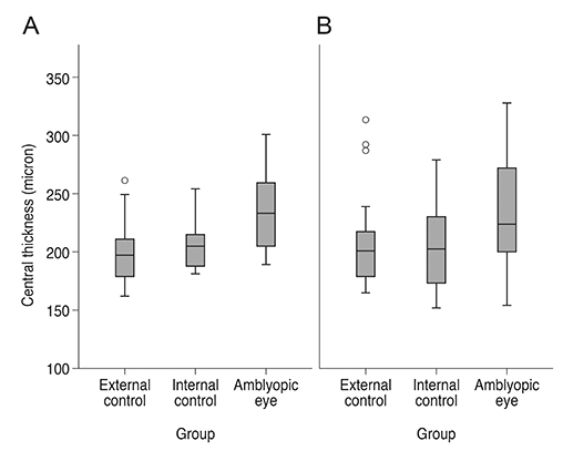
Fig. 1 Central retinal thickness (foveola) in (A) strabismic and (B) anisometropic amblyopic and control eyes.
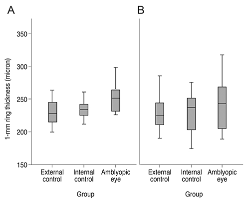
Fig. 2 Retinal thickness at 1-mm ring (fovea) in (A) strabismic and (B) anisometropic amblyopic and control eyes.
Reference
-
1. Liu H, Zhong L, Zhou X, Jin QZ. Macular abnormality observed by OCT in children with amblyopia failing to achieve normal visual acuity after long-term treatment. J Pediatr Ophthalmol Strabismus. 2010; 47:17–23.2. Kee SY, Lee SY, Lee YC. Thicknesses of the fovea and retinal nerve fiber layer in amblyopic and normal eyes in children. Korean J Ophthalmol. 2006; 20:177–181.
Article3. Altintas O, Yuksel N, Ozkan B, Caglar Y. Thickness of the retinal nerve fiber layer, macular thickness, and macular volume in patients with strabismic amblyopia. J Pediatr Ophthalmol Strabismus. 2005; 42:216–221.
Article4. Yoon SW, Park WH, Baek SH, Kong SM. Thicknesses of macular retinal layer and peripapillary retinal nerve fiber layer in patients with hyperopic anisometropic amblyopia. Korean J Ophthalmol. 2005; 19:62–67.
Article5. Yen MY, Cheng CY, Wang AG. Retinal nerve fiber layer thickness in unilateral amblyopia. Invest Ophthalmol Vis Sci. 2004; 45:2224–2230.
Article6. Bozkurt B, Irkec M, Orhan M, Karaagaoglu E. Thickness of the retinal nerve fiber layer in patients with anisometropic and strabismic amblyopia. Strabismus. 2003; 11:1–7.
Article7. Repka MX, Goldenberg-Cohen N, Edwards AR. Retinal nerve fiber layer thickness in amblyopic eyes. Am J Ophthalmol. 2006; 142:247–251.
Article8. Rajavi Z, Sabbaghi H, Baghini AS, et al. Prevalence of amblyopia and refractive errors among primary school children. J Ophthalmic Vis Res. 2015; 10:408–416.
Article9. Birch EE. Amblyopia and binocular vision. Prog Retin Eye Res. 2013; 33:67–84.
Article10. Altindag S. Evaluation of the macular thickness by optical coherence tomography in amblyopia. J Clin Exp Invest. 2016; 7:178–183.
Article11. Li J, Ji P, Yu M. Meta-analysis of retinal changes in unilateral amblyopia using optical coherence tomography. Eur J Ophthalmol. 2015; 25:400–409.
Article12. Kasem MA, Badawi AE. Changes in macular parameters in different types of amblyopia: optical coherence tomography study. Clin Ophthalmol. 2017; 11:1407–1416.13. Chen W, Xu J, Zhou J, et al. Thickness of retinal layers in the foveas of children with anisometropic amblyopia. PLoS One. 2017; 12:e0174537.
Article14. Rajavi Z, Moghadasifar H, Feizi M, et al. Macular thickness and amblyopia. J Ophthalmic Vis Res. 2014; 9:478–483.
Article15. Schalij-Delfos NE, de Graaf ME, Treffers WF, et al. Long term follow up of premature infants: detection of strabismus, amblyopia, and refractive errors. Br J Ophthalmol. 2000; 84:963–967.
Article16. West S, Williams C. Amblyopia. BMJ Clin Evid. 2011; 2011:0709.17. Amiri MA, Khosravi B, Far SH, et al. A comparative survey of OCT finding in fovea of unilateral persistent anisometropia amblyopic and normal fellow eyes. Sci J Rehabil Med. 2013; 2:1–10.18. Andalib D, Javadzadeh A, Nabai R, Amizadeh Y. Macular and retinal nerve fiber layer thickness in unilateral anisometropic or strabismic amblyopia. J Pediatr Ophthalmol Strabismus. 2013; 50:218–221.
Article19. Aguirre F, Mengual E, Hueso JR, Moya M. Comparison of normal and amblyopic retinas by optical coherence tomography in children. Eur J Ophthalmol. 2010; 20:410–418.
Article20. Kara O, Altintas O, Karaman S, et al. Analysis of choroidal thickness using spectral-domain OCT in children with unilateral amblyopia. J Pediatr Ophthalmol Strabismus. 2015; 52:159–166.
Article21. Al-Haddad C, Fattah MA, Ismail K, Bashshur Z. Choroidal changes in anisometropic and strabismic children with unilateral amblyopia. Ophthalmic Surg Lasers Imaging Retina. 2016; 47:900–907.
Article22. Liu Y, Dong Y, Zhao K. A meta-analysis of choroidal thickness changes in unilateral amblyopia. J Ophthalmol. 2017; 2017:2915261.
Article23. Ohno-Matsui K, Akiba M, Moriyama M, et al. Intrachoroidal cavitation in macular area of eyes with pathologic myopia. Am J Ophthalmol. 2012; 154:382–393.
Article
- Full Text Links
-
- Actions
-
Cited
- CITED
-
- Close
- Share
-
- Similar articles
-
- The Analysis of Peripapillary RNFL, Macula and Macular Ganglion Cell Layer Thickness in Patients with Monocular Amblyopia Using SD-OCT
- Thicknesses of Macular Retinal Layer and Peripapillary Retinal Nerve Fiber Layer in Patients with Hyperopic Anisometropic Amblyopia
- Efficacy of Occlusion Therapy in Amblyopia: Type, Depth and Timing of Amblyopia
- Macular Thickness and Visual Acuity Before and After Panretinal Photocoagulation in Severe Diabetic Retinopathy
- Diurnal Variation of Macular Thickness in Diabetic Macular Edema
