Effectiveness of Intravitreal Ranibizumab for Diabetic Macular Edema with Serous Retinal Detachment
- Affiliations
-
- 1Department of Ophthalmology, Dokuz Eylul University School of Medicine, Izmir, Turkey. mahmutkaya78@yahoo.com
- 2Department of Ophthalmology, Van Training and Research Hospital, Van, Turkey.
- KMID: 2418150
- DOI: http://doi.org/10.3341/kjo.2017.0117
Abstract
- PURPOSE
To evaluate the effectiveness of intravitreal injection of ranibizumab (IVR) in treating diabetic macular edema (DME) with serous retinal detachment (SRD) based on spectral domain optical coherence tomography (SD-OCT) patterns.
METHODS
One hundred thirty-four eyes of 134 patients with DME who underwent SD-OCT evaluation were included in this study. We retrospectively analyzed the medical records of patients who received IVR for the treatment of DME. Their eyes were classified into three groups according to the following SD-OCT features: SRD, diffuse retinal thickness and cystoid macular edema. The three groups were compared regarding changes in best-corrected visual acuity and central foveal thickness (CFT) after IVR.
RESULTS
The mean age was 61.4 ± 9.2 years (range, 44 to 81 years). The average length of the follow-up period was 9.4 ± 3.4 months (range, 6 to 24 months). The mean CFT value was significantly reduced in all groups (p < 0.001) after treatment. Increases in best-corrected visual acuity were statistically significant for the diffuse retinal thickness and cystoid macular edema groups (p < 0.001 and p < 0.001, respectively). However, there was no significant improvement after IVR injection in the SRD group (p = 0.252). In the SRD group, patients with ellipsoid zone disruption and external limiting membrane disruption demonstrated poorer visual gains at the last follow-up visit (p < 0.005 and p = 0.002, respectively).
CONCLUSIONS
A significant reduction in CFT with required IVR injections in DME with SRD was achieved but was accompanied by a worse functional outcome in the SRD group. The presence of subretinal fluid on SD-OCT in study eyes may be a poor prognostic factor for visual acuity.
Keyword
MeSH Terms
Figure
-
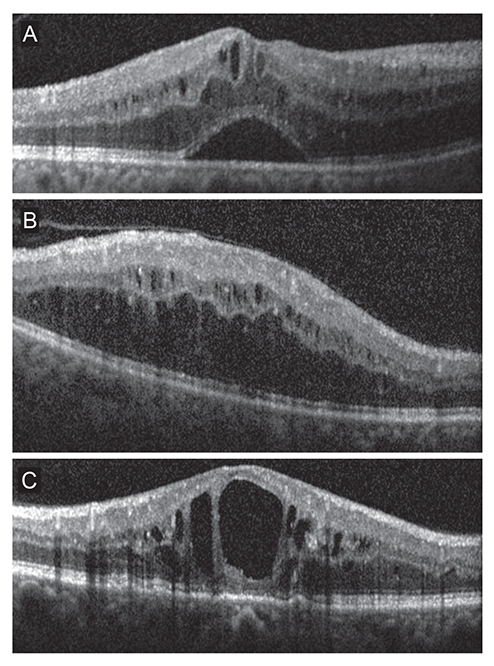
Fig. 1 Diabetic macular edema (DME) classification based on spectral domain optical coherence tomography. (A) Patients with predominant serous retinal detachment DME had an associated subretinal collection of fluid under the fovea. (B) Diffuse DME patients demonstrated widespread retinal thickening with a sponge-like appearance of the macula. (C) Patients with focal cystoid DME had a mound-like appearance of the fovea due to focal collection of fluid at the fovea.
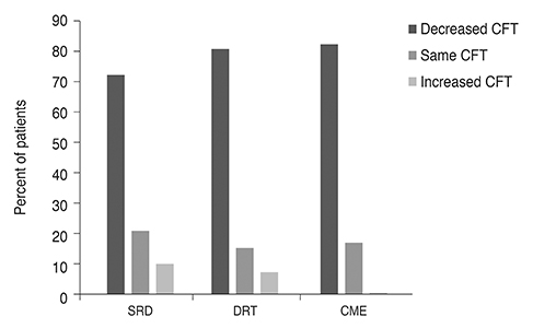
Fig. 2 Central foveal thickness (CFT) changes of the groups. SRD = serous retinal detachment; DRT = diffuse retinal thickness; CME = cystoid macular edema.
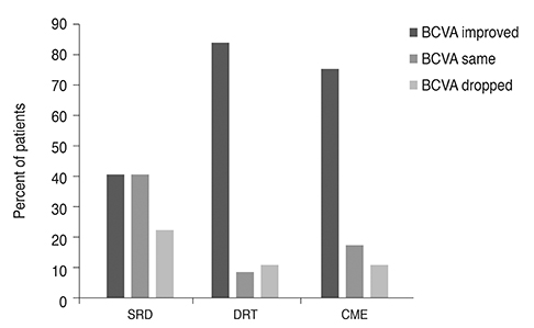
Fig. 3 Results of best-corrected visual acuity (BCVA) in eyes with decreased diabetic macular edema. SRD = serous retinal detachment; DRT = diffuse retinal thickness; CME = cystoid macular edema.
Reference
-
1. Patz A, Schatz H, Berkow JW, et al. Macular edema: an overlooked complication of diabetic retinopathy. Trans Am Acad Ophthalmol Otolaryngol. 1973; 77:OP34–OP42.2. Do Carmo A, Ramos P, Reis A, et al. Breakdown of the inner and outer blood retinal barrier in streptozotocin-induced diabetes. Exp Eye Res. 1998; 67:569–575.
Article3. Sander B, Larsen M, Moldow B, Lund-Andersen H. Diabetic macular edema: passive and active transport of fluorescein through the blood-retina barrier. Invest Ophthalmol Vis Sci. 2001; 42:433–438.4. Tso MO, Cunha-Vaz JG, Shih CY, Jones CW. Clinicopathologic study of blood-retinal barrier in experimental diabetes mellitus. Arch Ophthalmol. 1980; 98:2032–2040.
Article5. Bhagat N, Grigorian RA, Tutela A, Zarbin MA. Diabetic macular edema: pathogenesis and treatment. Surv Ophthalmol. 2009; 54:1–32.
Article6. Otani T, Kishi S, Maruyama Y. Patterns of diabetic macular edema with optical coherence tomography. Am J Ophthalmol. 1999; 127:688–693.
Article7. Kang SW, Park CY, Ham DI. The correlation between fluorescein angiographic and optical coherence tomographic features in clinically significant diabetic macular edema. Am J Ophthalmol. 2004; 137:313–322.
Article8. Ciulla TA, Harris A, Latkany P, et al. Ocular perfusion abnormalities in diabetes. Acta Ophthalmol Scand. 2002; 80:468–477.
Article9. Thomas BJ, Shienbaum G, Boyer DS, Flynn HW Jr. Evolving strategies in the management of diabetic macular edema: clinical trials and current management. Can J Ophthalmol. 2013; 48:22–30.
Article10. Stewart MW. Anti-vascular endothelial growth factor drug treatment of diabetic macular edema: the evolution continues. Curr Diabetes Rev. 2012; 8:237–246.11. Massin P, Bandello F, Garweg JG, et al. Safety and efficacy of ranibizumab in diabetic macular edema (RESOLVE Study): a 12-month, randomized, controlled, double-masked, multicenter phase II study. Diabetes Care. 2010; 33:2399–2405.
Article12. Diabetic Retinopathy Clinical Research Network. Scott IU, Edwards AR, et al. A phase II randomized clinical trial of intravitreal bevacizumab for diabetic macular edema. Ophthalmology. 2007; 114:1860–1867.
Article13. Arevalo JF, Sanchez JG, Wu L, et al. Primary intravitreal bevacizumab for diffuse diabetic macular edema: the Pan-American Collaborative Retina Study Group at 24 months. Ophthalmology. 2009; 116:1488–1497.14. Koytak A, Altinisik M, Sogutlu Sari E, et al. Effect of a single intravitreal bevacizumab injection on different optical coherence tomographic patterns of diabetic macular oedema. Eye (Lond). 2013; 27:716–721.
Article15. Cheema HR, Al Habash A, Al-Askar E. Improvement of visual acuity based on optical coherence tomography patterns following intravitreal bevacizumab treatment in patients with diabetic macular edema. Int J Ophthalmol. 2014; 7:251–255.16. Petrella RJ, Blouin J, Davies B, Barbeau M. Prevalence, demographics, and treatment characteristics of visual impairment due to diabetic macular edema in a representative Canadian cohort. J Ophthalmol. 2012; 2012:159167.
Article17. Kim M, Lee P, Kim Y, et al. Effect of intravitreal bevacizumab based on optical coherence tomography patterns of diabetic macular edema. Ophthalmologica. 2011; 226:138–144.
Article18. Wu PC, Lai CH, Chen CL, Kuo CN. Optical coherence tomographic patterns in diabetic macula edema can predict the effects of intravitreal bevacizumab injection as primary treatment. J Ocul Pharmacol Ther. 2012; 28:59–64.
Article19. Shimura M, Yasuda K, Yasuda M, Nakazawa T. Visual outcome after intravitreal bevacizumab depends on the optical coherence tomographic patterns of patients with diffuse diabetic macular edema. Retina. 2013; 33:740–747.
Article20. Seo KH, Yu SY, Kim M, Kwak HW. Visual and morphologic outcomes of intravitreal ranibizumab for diabetic macular edema based on optical coherence tomography patterns. Retina. 2016; 36:588–595.
Article21. Giocanti-Auregan A, Hrarat L, Qu LM, et al. Functional and anatomical outcomes in patients with serous retinal detachment in diabetic macular edema treated with ranibizumab. Invest Ophthalmol Vis Sci. 2017; 58:797–800.22. Antonetti DA, Barber AJ, Khin S, et al. Vascular permeability in experimental diabetes is associated with reduced endothelial occludin content: vascular endothelial growth factor decreases occludin in retinal endothelial cells. Penn State Retina Research Group. Diabetes. 1998; 47:1953–1959.
Article23. Aiello LP, Avery RL, Arrigg PG, et al. Vascular endothelial growth factor in ocular fluid of patients with diabetic retinopathy and other retinal disorders. N Engl J Med. 1994; 331:1480–1487.
Article24. Neufeld G, Cohen T, Gengrinovitch S, Poltorak Z. Vascular endothelial growth factor (VEGF) and its receptors. FASEB J. 1999; 13:9–22.
Article25. Grant MB, Afzal A, Spoerri P, et al. The role of growth factors in the pathogenesis of diabetic retinopathy. Expert Opin Investig Drugs. 2004; 13:1275–1293.
Article26. Gaucher D, Sebah C, Erginay A, et al. Optical coherence tomography features during the evolution of serous retinal detachment in patients with diabetic macular edema. Am J Ophthalmol. 2008; 145:289–296.
Article27. Ozdemir H, Karacorlu M, Karacorlu S. Serous macular detachment in diabetic cystoid macular oedema. Acta Ophthalmol Scand. 2005; 83:63–66.
Article28. Fatt I, Shantinath K. Flow conductivity of retina and its role in retinal adhesion. Exp Eye Res. 1971; 12:218–226.
Article29. Nagelhus EA, Veruki ML, Torp R, et al. Aquaporin-4 water channel protein in the rat retina and optic nerve: polarized expression in Müller cells and fibrous astrocytes. J Neurosci. 1998; 18:2506–2519.
Article
- Full Text Links
-
- Actions
-
Cited
- CITED
-
- Close
- Share
-
- Similar articles
-
- Long-term Outcomes of Diabetic Macular Edema Following Initial Intravitreal Ranibizumab Injection Based on Morphologic Pattern
- The Short-Term Efficacy of Intravitreal Ranibizumab in the Treatment of Diabetic Macular Edema
- A Multimodal Approach to Diabetic Macular Edema
- The Short-term Treatment Efficacy of Intravitreal Ranibizumab Injection in TreatmentNaïve Patients with Diabetic Macular Edema
- Patterns of Macular Edema in Patients with Branch Retinal Vein Occlusion on Optical Coherence Tomography
