Comparison of Ocular Biometry Using New Swept-source Optical Coherence Tomography-based Optical Biometer with Other Devices
- Affiliations
-
- 1Department of Ophthalmology, HanGil Eye Hospital, Incheon, Korea. chobjn@empas.com
- 2Department of Ophthalmology, Catholic Kwandong University College of Medicine, Gangeung, Korea.
- KMID: 2418145
- DOI: http://doi.org/10.3341/kjo.2017.0091
Abstract
- PURPOSE
To evaluate the agreement between optical biometry with swept-source optical coherence tomography-based optical biometry (IOLMaster 700) and other devices.
METHODS
A total of 137 eyes (78 patients) with cataracts were included in this retrospective study. Axial length (AL), anterior chamber depth (ACD), keratometry, and white-to-white (WTW) distance measured using IOLMaster 700 were compared with results for the following five different biometers: IOLMaster 500, A-scan, automated refractor, manual keratometry, and Galilei G4. Differences and correlations among the devices were assessed using the Bland-Altman plot and intraclass correlation coefficient (ICC).
RESULTS
For AL values, the IOLMaster 700, IOLMaster 500, and A-scan measurements showed excellent agreement (all ICC >0.99). For ACD values, ICC of IOLMaster 700 and Galilei G4 was 0.965 but A-scan was poorly correlated with either IOLMaster 700 or Galilei G4. The ICCs of IOLMaster 700 and other devices were all greater than 0.9 for average keratometry, but those of the mean cylinder keratometry were all between 0.7 and 0.8. The mean difference in the WTW distance between the IOLMaster 700 and Galilei G4 was 0.029 mm, but the ICC was 0.525. AL measurements were not possible for 10 eyes with the IOLMaster 500 but were obtained in all eyes with the IOLMaster 700.
CONCLUSIONS
In clinical practice, AL, ACD, and average keratometry values of IOLMaster 700 can be used interchangeably with those of the other devices tested. However, the ACD value between IOLMaster 700 and A-scan or the WTW distance between IOLMaster 700 and Galilei G4 are not interchangeable because of clinical and statistical differences in measurements between the devices.
Keyword
Figure
-

Fig. 1 Bland-Altman plot showing agreements in axial length (mm) measurements. (A) IOLMaster 700 and IOLMaster 500, (B) IOLMaster 700 and A-scan, and (C) IOLMaster 500 and A-scan. The solid line represents the mean difference and the dotted lines represent the 95% limits of agreement.

Fig. 2 Bland-Altman plot showing the agreements in anterior chamber depth (mm) measurements. (A) IOLMaster 700 and Galilei G4, (B) IOLMaster 700 and A-scan, and (C) Galilei G4 and A-scan. The solid line represents the mean difference and the dotted lines represent the 95% limits of agreement.

Fig. 3 Bland-Altman plots of the agreements in mean keratometry (diopter) measurements. (A) IOLMaster 700 and IOLMaster 500, (B) IOLMaster 700 and Galilei G4, (C) IOLMaster 700 and automated refractor, and (D) IOLMaster 700 and manual keratometry. The solid line represents the mean difference and the dotted lines represent the 95% limits of agreement.
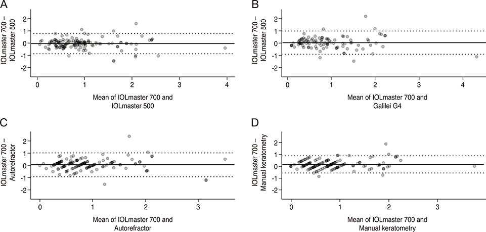
Fig. 4 Bland-Altman plots of the agreements in mean cylinder power (diopter) measurements. (A) IOLMaster 700 and IOLMaster 500, (B) IOLMaster 700 and Galilei G4, (C) IOLMaster 700 and automated refractor, and (D) IOLMaster 700 and manual keratometry. The solid line represents the mean difference and the dotted lines represent the 95% limits of agreement.
Reference
-
1. Srivannaboon S, Chirapapaisan C, Chonpimai P, Loket S. Clinical comparison of a new swept-source optical coherence tomography-based optical biometer and a time-domain optical coherence tomography-based optical biometer. J Cataract Refract Surg. 2015; 41:2224–2232.
Article2. Akman A, Asena L, Gungor SG. Evaluation and comparison of the new swept source OCT-based IOLMaster 700 with the IOLMaster 500. Br J Ophthalmol. 2016; 100:1201–1205.
Article3. Hoffer KJ, Hoffmann PC, Savini G. Comparison of a new optical biometer using swept-source optical coherence tomography and a biometer using optical low-coherence reflectometry. J Cataract Refract Surg. 2016; 42:1165–1172.
Article4. Landis JR, Koch GG. The measurement of observer agreement for categorical data. Biometrics. 1977; 33:159–174.
Article5. Stopyra W. The accuracy of IOL power calculation formulas for eyes of axial length exceeding 24.5 mm. Klin Oczna. 2013; 115:93–95.6. Salouti R, Nowroozzadeh MH, Zamani M, et al. Comparison of the ultrasonographic method with 2 partial coherence interferometry methods for intraocular lens power calculation. Optometry. 2011; 82:140–147.
Article7. Kunert KS, Peter M, Blum M, et al. Repeatability and agreement in optical biometry of a new swept-source optical coherence tomography-based biometer versus partial coherence interferometry and optical low-coherence reflectometry. J Cataract Refract Surg. 2016; 42:76–83.
Article8. Yang JY, Kim HK, Kim SS. Axial length measurements: comparison of a new swept-source optical coherence tomography-based biometer and partial coherence interferometry in myopia. J Cataract Refract Surg. 2017; 43:328–332.
Article9. Gursoy H, Sahin A, Basmak H, et al. Lenstar versus ultrasound for ocular biometry in a pediatric population. Optom Vis Sci. 2011; 88:912–919.
Article10. Sanchis-Gimeno JA, Palanca-Sanfrancisco JM, Garcia-Lazaro S, et al. The effect of anesthetic eye drop instillation on the distribution of corneal thickness. Cornea. 2013; 32:e102–e105.
Article11. Landers J, Goggin M. Comparison of refractive outcomes using immersion ultrasound biometry and IOLMaster biometry. Clin Exp Ophthalmol. 2009; 37:566–569.
Article12. Patel RP, Pandit RT. Comparison of anterior chamber depth measurements from the Galilei dual Scheimpflug analyzer with IOLMaster. J Ophthalmol. 2012; 2012:430249.
Article13. Elbaz U, Barkana Y, Gerber Y, et al. Comparison of different techniques of anterior chamber depth and keratometric measurements. Am J Ophthalmol. 2007; 143:48–53.
Article14. Reddy AR, Pande MV, Finn P, El-Gogary H. Comparative estimation of anterior chamber depth by ultrasonography, Orbscan II, and IOLMaster. J Cataract Refract Surg. 2004; 30:1268–1271.
Article15. Santodomingo-Rubido J, Mallen EA, Gilmartin B, Wolffsohn JS. A new non-contact optical device for ocular biometry. Br J Ophthalmol. 2002; 86:458–462.
Article16. Hashemi H, Yazdani K, Mehravaran S, Fotouhi A. Anterior chamber depth measurement with a-scan ultrasonography, Orbscan II, and IOLMaster. Optom Vis Sci. 2005; 82:900–904.17. Dominguez-Vicent A, Perez-Vives C, Ferrer-Blasco T, et al. Device interchangeability on anterior chamber depth and white-to-white measurements: a thorough literature review. Int J Ophthalmol. 2016; 9:1057–1065.
Article18. Whang WJ, Byun YS, Joo CK. Comparison of refractive outcomes using five devices for the assessment of preoperative corneal power. Clin Exp Ophthalmol. 2012; 40:425–432.
Article19. Shirayama M, Wang L, Weikert MP, Koch DD. Comparison of corneal powers obtained from 4 different devices. Am J Ophthalmol. 2009; 148:528–535.
Article
- Full Text Links
-
- Actions
-
Cited
- CITED
-
- Close
- Share
-
- Similar articles
-
- Comparison of Clinical Outcomes between Swept-source Optical Coherence Tomography Biometer and Partial Coherence Interferometer
- Accuracy of Predicting Refractive Outcomes Using Swept-source Optical Coherence Tomography in Nuclear Cataracts
- Comparison of Ocular Biometric Measurements Using New Swept-source Optical-coherence Tomography, Low-coherence Reflectometry, A-Scan Biometry, and Autokeratometry
- Comparison of Image Quality between Swept-Source and Spectral-Domain Optical Coherence Tomography According to Ocular Media Opacity
- Comparison of Anterior Segment Measurements Between Swept-source Optical Coherence Tomography and Schiempflug Coherence Interferometer
