The Availability of Quantitative Assessment of Pain Perception in Patients With Diabetic Polyneuropathy
- Affiliations
-
- 1Department of Rehabilitation Medicine, Yonsei University Wonju College of Medicine, Wonju, Korea. lighthouse14@naver.com
- KMID: 2417835
- DOI: http://doi.org/10.5535/arm.2018.42.3.433
Abstract
OBJECTIVE
To evaluate the usefulness of the quantitative assessment of pain perception (QAPP) in diabetic polyneuropathy (DPN) patients.
METHODS
Thirty-two subjects with DPN were enrolled in this study. The subjects' pain perception was assessed quantitatively. Current perception threshold (CPT) and pain equivalent current (PEC) were recorded. All patients were tested with a nerve conduction study (NCS) for evaluation of DPN and pain-related evoked potential (PREP) for evaluation of small fiber neuropathy (SFN) on bilateral upper and lower limbs. All patients were asked to participate in tests such as visual analogue scale (VAS) and SF-36 Health Survey Version 2 to evaluate their subjective pain and quality of life, respectively.
RESULTS
The PEC of QAPP showed significant correlations with VAS (p=0.002) and physical function surveyed with SF-36 Health Survey Version 2 (p=0.035). The results of QAPP had no correlation with NCS, but there was a significant relationship between the CPT of QAPP and PREP (p=0.003).
CONCLUSION
The QAPP may be useful not only in providing objective evaluations of subjective pain in patients with DPN but also in the assessment of diabetic SFN.
MeSH Terms
Figure
-
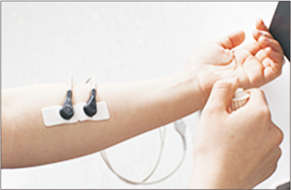
Fig. 1. Measuring position for quantitative assessment of pain perception. While in a comfortable seating position, the electrode patch is attached to the left anterior forearm and the stop switch is held in the right hand.

Fig. 2. (A) PEC of QAPP and pain degree showed significant positive correlation (correlation coefficient=0.956, p<0.0001). (B) PEC of QAPP and VAS showed significant positive correlation (correlation coefficient=0.526, p=0.002). (C) PEC of QAPP and physical function in SF-36v2 showed significant negative correlation (correlation coefficient=-0.373, p=0.035). (D) CPT of QAPP and P latency of PREP which was performed in C7 dermatome on the left side showed significant negative correlation (correlation coefficient=-0.511, p=0.003). PEC, pain equivalent current; QAPP, quantitative assessment of pain perception; VAS, visual analogue scale (0–100); CPT, current perception threshold; PREP, pain-related evoked potential.
Reference
-
1. Aguiree F, Brown A, Cho NH, Dahlquist G, Dodd S, Dunning T, et al. IDF diabetes atlas. 6th ed. Basel: International Diabetes Federation;2013.2. Redmond JM, McKenna MJ, Feingold M, Ahmad BK. Sensory testing versus nerve conduction velocity in diabetic polyneuropathy. Muscle Nerve. 1992; 15:1334–9.
Article3. Joo IS. Controversies on the usefulness of nerve conduction study in the early diagnosis of diabetic polyneuropathy. Ann Clin Neurophysiol. 2008; 10:25–8.4. American Diabetes Association American Academy of Neurology. Consensus statement: report and recommendations of the San Antonio conference on diabetic neuropathy. Diabetes Care. 1988; 11:592–7.5. Park JM, Kang SJ, Kim KW, Kim JW, Kim SH. Evaluation of peripheral polyneuropathy in patients with diabetes mellitus using quantitative sensory test. J Korean Acad Rehabil Med. 2001; 25:102–9.6. Mueller D, Obermann M, Koeppen S, Kavuk I, Yoon MS, Sack F, et al. Electrically evoked nociceptive potentials for early detection of diabetic small-fiber neuropathy. Eur J Neurol. 2010; 17:834–41.
Article7. Bradham DD. Outcomes research in orthopedics: history, perspectives, concepts, and future. Arthroscopy. 1994; 10:493–501.
Article8. Herr KA, Mobily PR, Kohout FJ, Wagenaar D. Evaluation of the Faces Pain Scale for use with the elderly. Clin J Pain. 1998; 14:29–38.
Article9. Hicks CL, von Baeyer CL, Spafford PA, van Korlaar I, Goodenough B. The Faces Pain Scale-Revised: toward a common metric in pediatric pain measurement. Pain. 2001; 93:173–83.10. Johnson LL, Pittsley A, Becker R, Young AD. A novel quantitative pain assessment instrument that provides means of comparing patient’s pain magnitude with a measurement of their pain tolerance. J Clin Med Res. 2015; 7:781–90.
Article11. Hansson P, Backonja M, Bouhassira D. Usefulness and limitations of quantitative sensory testing: clinical and research application in neuropathic pain states. Pain. 2007; 129:256–9.
Article12. Ohtori S, Kawaguchi H, Takebayashi T, Orita S, Inoue G, Yamauchi K, et al. PainVision apparatus is effective for assessing low back pain. Asian Spine J. 2014; 8:793–8.
Article13. Chun SW, Ko KS. Summary of the update to the diabetic neuropathy management guidebook. J Korean Diabetes. 2012; 13:115–23.
Article14. Jin WJ, Yu TY, Jin YH, Lee JB. Visual analogue scale in acute pain measurement: its usefulness as a pain measurement tool in an emergency setting. J Korean Soc Emerg Med. 2003; 14:61–5.15. Bijur PE, Silver W, Gallagher EJ. Reliability of the visual analog scale for measurement of acute pain. Acad Emerg Med. 2001; 8:1153–7.
Article16. Jensen MP, McFarland CA. Increasing the reliability and validity of pain intensity measurement in chronic pain patients. Pain. 1993; 55:195–203.
Article17. Jenkinson C, Wright L, Coulter A. Criterion validity and reliability of the SF-36 in a population sample. Qual Life Res. 1994; 3:7–12.
Article18. Tesfaye S, Boulton AJ, Dyck PJ, Freeman R, Horowitz M, Kempler P, et al. Diabetic neuropathies: update on definitions, diagnostic criteria, estimation of severity, and treatments. Diabetes Care. 2010; 33:2285–93.
Article19. Lee SM, Kim BJ. Diagnostic usefulness of quantitative sensory test in diabetic polyneuropathy: comparison with nerve conduction study. J Korean Neurol Assoc. 1999; 17:106–11.20. Donaghue VM, Giurini JM, Rosenblum BI, Weissman PN, Veves A. Variability in function measurements of three sensory foot nerves in neuropathic diabetic patients. Diabetes Res Clin Pract. 1995; 29:37–42.
Article21. Borsey DQ, Cull RE, Fraser DM, Ewing DJ, Campbell IW, Clarke BF. Small muscle wasting of the hands in diabetes mellitus. Diabetes Care. 1983; 6:10–7.
Article22. Ziegler D, Mayer P, Wiefels K, Gries FA. Assessment of small and large fiber function in long-term type 1 (insulin-dependent) diabetic patients with and without painful neuropathy. Pain. 1988; 34:1–10.
Article23. Jamal GA, Hansen S, Weir AI, Ballantyne JP. The neurophysiologic investigation of small fiber neuropathies. Muscle Nerve. 1987; 10:537–45.
Article24. Dyck PJ, Lambert EH, O’Brien PC. Pain in peripheral neuropathy related to rate and kind of fiber degeneration. Neurology. 1976; 26:466–71.
Article25. Cazzato D, Lauria G. Small fibre neuropathy. Curr Opin Neurol. 2017; 30:490–9.
Article26. Hovaguimian A, Gibbons CH. Diagnosis and treatment of pain in small-fiber neuropathy. Curr Pain Headache Rep. 2011; 15:193–200.
Article27. Katsarava Z, Ayzenberg I, Sack F, Limmroth V, Diener HC, Kaube H. A novel method of eliciting pain-related potentials by transcutaneous electrical stimulation. Headache. 2006; 46:1511–7.
Article
- Full Text Links
-
- Actions
-
Cited
- CITED
-
- Close
- Share
-
- Similar articles
-
- Evaluation of Peripheral Polyneuropathy in Patients with Diabetes Mellitus Using Quantitative Sensory Test
- Clinical Evaluation for Diabetic Neuropathy
- Correlation Between the Severity of Diabetic Peripheral Polyneuropathy and Glycosylated Hemoglobin Levels: A Quantitative Study
- Usefulness of Questionnaires, Physical Examination and Median Mixed Nerve Conduction Studies in Patients with Diabetes Mellitus
- Diagnostic Usefulness of Quantitative Sensory Test in Diabetic Polyneuropathy: Comparison with Nerve Conduction Study
