Hepatobiliary diseases in buffalo (Bubalus bubalis): clinical, laboratory, and ultrasonographic findings
- Affiliations
-
- 1Division of Internal Medicine, Department of Animal Medicine, Assiut University, Assiut 71526, Egypt. arafatvet2003@yahoo.com
- 2Department of Surgery, Anesthesiology and Radiology, Faculty of Veterinary Medicine, Cairo University, Giza 12211, Egypt.
- 3Veterinary Teaching Hospital, Faculty of Veterinary Medicine, Assiut University, Assiut 71526, Egypt.
- 4Division of Clinical Laboratory Diagnosis, Department of Animal Medicine, Assiut University, Assiut 71526, Egypt.
- 5Department of Surgery, Anaesthesiology and Radiology, Faculty of Veterinary Medicine, Aswan University, Aswan 81528, Egypt.
- 6Division of Clinical Laboratory Diagnosis, Department of Animal Medicine, Faculty of Veterinary Medicine, Sohag University, Sohag 82524, Egypt.
- KMID: 2417570
- DOI: http://doi.org/10.4142/jvs.2018.19.4.543
Abstract
- This study describes ultrasonographic observations of five hepatobiliary diseases in buffalo (Bubalus bubalis). Fifty buffalo, including 20 clinically normal and 30 hepatobiliary diseased buffalo were enrolled in the study. Complete clinical, radiographic and ultrasonographic examinations and laboratory analyses were conducted. Focal parenchymal lesions including liver abscess (n = 12) and hepatic cyst (n = 6), diffuse parenchymal lesion (hepatobiliary cirrhosis, n = 5) and obstruction of hepatobiliary passages including cholestasis (n = 4), and hepatocholelithiasis (n = 3) were successfully imaged by ultrasonography. Hepatic abscess imaged as a hypoechoic to echogenic circumscribed mass of various diameters with a distinct echogenic capsule. Hepatic cyst imaged as a pear-shaped sac with a bright echogenic margin, anechoic content, and distal acoustic enhancement. In hepatobiliary fibrosis, the liver showed linear bands of increasing echogenicity with less distinct imaging of the portal vasculature. Cholestasis was imaged as dilatation of the gallbladder (GB) with wall thickening and homogeneous or heterogeneous contents. Hepatocholelithiasis imaged as an echoic structure within the hepatic parenchyma, or within and around the GB and bile duct, with more echogenicity of the hepatic parenchyma than normal. Ultrasonography can be an efficient rapid, noninvasive tool for screening of common hepatobiliary diseases in buffalo under field conditions.
Keyword
MeSH Terms
Figure
-
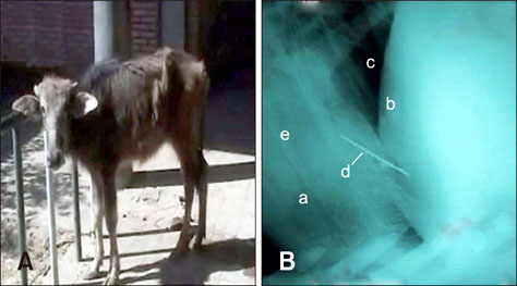
Fig. 1 (A) A buffalo heifer with traumatic reticuloperitonitis and hepatic abscess and stunted growth. (B) Lateral radiograph of the cranial abdomen and caudal thorax of a 5-year-old non-pregnant buffalo with traumatic pericarditis showing a nail passing from the reticulum and penetrating the heart. a, heart; b, reticulum; c, diaphragm; d, nail; e, loss of heart details.
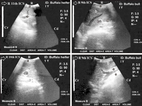
Fig. 2 Ultrasonograms of 1-year-old buffalo heifer (left) and 1-year-old buffalo bull (right) showing liver (b) with hepatic abscess (c) when imaged from the right 9th, 10th, and 11th intercostal space (ICS). Right abdominal wall (a). Liver abscess was imaged as well a distinct echogenic capsule and hypoechoic lumen. An echogenic clear tract (d) was imaged within the hepatic tissue with accumulation of echogenic deposits around the gallbladder and the abscess. The abscess formed a thick layer of echogenic deposits around the cystic duct as pericholecystic hyperechogenic material (e). R, right; Cr, cranial; Cd, caudal.

Fig. 3 Ultrasonogram in a 1-year-old non-pregnant buffalo heifer showing liver (b) with hepatic cyst (c) in an image from the right 11th intercostal space (ICS). Right abdominal wall (a). The cyst appears as a pear-shaped structure (black cyst) with bright echogenic margin and a well-defined thin wall located caudoventral to the liver and causing distal acoustic enhancement (d). R, right; Cr, cranial; Cd, caudal.
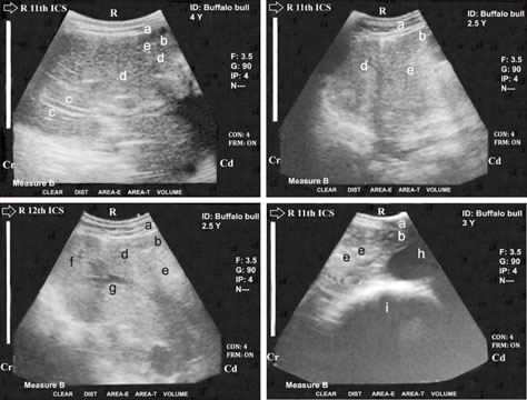
Fig. 4 Ultrasonogram in 2.5, 3, and 4-year-old buffalo bulls obtained at the right 11th and 12th intercostal space (ICS). Right abdominal wall (a). Image shows liver (b) fibrosis/cirrhosis and visualization of linear bands of increasing echogenicity (c) with less distinct visualization of the hepatic and portal vasculatures. Image shows multiple hyperechogenic foci (e) within the hepatic parenchyma with anechoic nodules (d) indicating the heterogeneous nature of the hepatic parenchyma. Reduced lumens of both of caudal vena cava (f) and hepatic vein (g) with accumulation of fibrinous echogenic deposits around the vein are present. Gallbladder (h) and cranial duodenum (i) are also visible. R, right; Cr, cranial; Cd, caudal.

Fig. 5 Ultrasonogram in a 1-year-old buffalo heifer with liver cirrhosis and dilated loops of the cranial duodenum. Images from the right 10th and 11th intercostal space (ICS). Right abdominal wall (a). Multiple hyperechogenic foci (b) located within the hepatic parenchyma with anechoic nodules (c) with a heterogeneous hepatic parenchyma and less distinct visualization of the hepatic and portal vasculatures. Reduced lumens of both of caudal vena cava (d) and hepatic vein with presence of accumulated fibrinous echogenic deposits around the vein. The dilated loop of the cranial part of the duodenum (6.2–6.5 cm; e) was intertangled medially to the gallbladder (f) and liver. R, right; Cr, cranial; Cd, caudal.
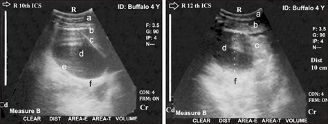
Fig. 6 Ultrasonogram in a 4-year-old non-pregnant buffalo imaged from the right 10th (left) and 12th (right) intercostal spaces (ICSs). Right abdominal wall (a). Image shows healthy liver tissue with normal parenchymal pattern (b) and a dilated gallbladder (cholestasis; c). The dilated gallbladder had a heterogeneous nature with echoic and hypoechoic contents (d), thickening in its wall (e) and distal acoustic enhancement (f). R, right; Cr, cranial; Cd, caudal.
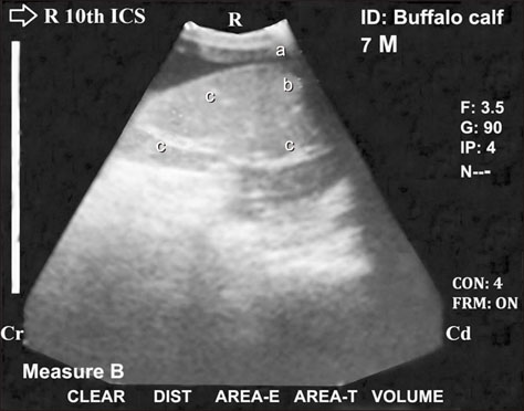
Fig. 7 Ultrasonogram in a 7-month-old fattening buffalo calf imaged from the right 10th intercostal space (ICS). Right abdominal wall (a). Image shows liver (b) with hepatocholelithiasis, which revealed hyperechogenic masses or deposits scattered (c) within the hepatic tissue along with increased hepatic parenchymal echogenicity when compared with normal parenchymal echogenicity of the liver. R, right; Cr, cranial; Cd, caudal.
Reference
-
1. Abdelaal AM, Gouda SM, Tharwat M. Clinico-biochemical, ultrasonographic and pathological findings of hepatic abscess in feedlot cattle and buffaloes. Vet World. 2014; 7:306–310.
Article2. Abdelaal AM, Mostafa MB, Abu-Seida AM, Al-Abbadi OS, Abbas SF. Ultrasonographic findings in hardware diseased buffaloes (Babulus babilus). Res J Pharmaceut Biol Chem Sci. 2016; 7:1644–1649.3. Abu-Seida AM. Current status and prospect of ultrasonographic application in buffaloes. Asian J Anim Vet Adv. 2016; 11:144–157.
Article4. Abu-Seida AM, Al-Abbadi OS. Recent advances in the management of foreign body syndrome in cattle and buffaloes: a review. Pak Vet J. 2016; 36:385–393.5. Abu-Seida AM, Al-Abbadi OS. Studies on sharp foreign body syndrome in Iraqi buffaloes and its impact on milk production. Asian J Anim Vet Adv. 2015; 9:128–133.
Article6. Al-Abbadi OS, Abu-Seida AM, Al-Hussainy SM. Studies on rumen magnet usage to prevent hardware disease in buffaloes. Vet World. 2014; 7:408–411.
Article7. Andrews AH, Blowey RW, Boyd H, Eddy RG. Bovine Medicine, Diseases and Husbandry of Cattle. 2nd ed. Ames: Blackwell Science;2004. p. 835–839.8. Berhe G. Abattoir survey on cattle hydatidosis in Tigray Region of Ethiopia. Trop Anim Health Prod. 2009; 41:1347–1352.
Article9. Blond L, Buczinski S. Basis of ultrasound imaging and the main artifacts in bovine medicine. Vet Clin North Am Food Anim Pract. 2009; 25:553–565.
Article10. Brandon B, Stanley C. What is your diagnosis? Cholestasis, hepatic cholelithiasis. J Am Vet Med Assoc. 2003; 222:289–290.11. Braun U. Ultrasonography of the liver in cattle. Vet Clin North Am Food Anim Pract. 2009; 25:591–609.
Article12. Braun U, Flückiger M, Nägeli F. Radiography as an aid in the diagnosis of traumatic reticuloperitonitis in cattle. Vet Rec. 1993; 132:103–109.
Article13. Braun U, Götz M. Ultrasonography of the reticulum in cows. Am J Vet Res. 1994; 55:325–332.14. Braun U, Marmier O, Pusterla N. Ultrasonographic examination of the small intestine of cows with ileus of the duodenum, jejunum or ileum. Vet Rec. 1995; 137:209–215.
Article15. Braun U, Pusterla N, Wild K. Ultrasonographic findings in 11 cows with a hepatic abscess. Vet Rec. 1995; 137:284–290.
Article16. Cockcroft PD. Diagnosis and clinical reasoning in cattle practice. In : Cockcroft PD, editor. Bovine Medicine. 3rd ed. Hoboken: John Wiley & Sons;2015. p. 124–132.17. Coles EH. Veterinary Clinical Pathology. 4th ed. Philadelphia: W.B. Saunders;1986. p. 132–139.18. Khalphallah A, Abdelhakiem M, Elmeligy E. The ultrasonographic findings of the liver, gall bladder and their related vasculatures in healthy Egyptian buffaloes (Bubalus bubalis). Assiut Vet Med J. 2016; 62:156–162.19. Khalphallah A, Abu-Seida AM, Abdelhakiem M, Elmeligy E, Mahmoud UT. Laboratory, radiographic and ultrasonographic findings of acute traumatic reticuloperitonitis in buffaloes (Bubalus bubalis). Asian J Anim Vet Adv. 2016; 11:675–683.20. Khalphallah A, Aref NE, Elmeligy E, El-Hawari SF. Clinical and ultrasonographic observations of functional and mechanical intestinal obstruction in buffaloes (Bubalus bubalis). Vet World. 2016; 9:475–480.
Article21. Khalphallah A, Elmeligy E, El-Hawari SF, Mahmoud UT. Clinical, laboratory and ultrasonographic findings in Egyptian buffalo (Bubalus bubalis) with caecal and colonic dilatation. Int J Vet Sci Med. 2016; 4:5–10.
Article22. Khalphallah A, Elmeligy E, Elsayed HK, El-Hawari SF, Elrashidy MH. Diagnostic significance of ultrasonography in complicated traumatic reticuloperitonitis in Egyptian buffaloes (Bubalus bubalis). Asian J Anim Vet Adv. 2016; 11:319–330.23. Khalphallah A, El-Sebaie A, Raghib M. Cows with heptaobiliary diseases: clinical, biochemical and ultrasonographic findings. Scholar's Adv Anim Vet Res. 2016; 3:56–67.24. Khalphallah AA, El-Sebaie AH, Raghib MF. Approach for diagnosis of complicated traumatic reticuloperitonitis in cattle using ultrasonography. J Adv Vet Res. 2015; 5:157–164.25. Lahmar S, Chéhida FB, Pétavy AF, Hammou A, Lahmar J, Ghannay A, Gharbi HA, Sarciron ME. Ultrasonographic screening for cystic echinococcosis in sheep in Tunisia. Vet Parasitol. 2007; 143:42–49.
Article26. Nagaraja TG, Lechtenberg KF. Liver abscesses in feedlot cattle. Vet Clin North Am Food Anim Pract. 2007; 23:351–369.
Article27. Radostis OM, Gaym CC, Hinchcliff KW, Constable PD. Veterinary Medicine. A Textbook of The Diseases of Cattle, Horses, Sheep, Pigs and Goats. 10th ed. Philadelphia: Saunders Elsevier;2007. p. 189–138.28. Tennant BC. Hepatic function. In : Kaneko JJ, Harvey JW, Bruss ML, editors. Clinical Biochemistry of Domestic Animals. 6th ed. New York: Academic Press;2008. p. 379–341.29. Tesfaye D, Chanie M. Study on rumen and reticulum foreign bodies in cattle slaughtered at Jimma Municipal Abattoir, South West Ethiopia. Am-Euras J Sci Res. 2012; 7:160–167.
- Full Text Links
-
- Actions
-
Cited
- CITED
-
- Close
- Share
-
- Similar articles
-
- The use of crossreactive monoclonal antibodies to characterize the immune system of the water buffalo (Bubalus bubalis)
- Alginate encapsulation preserves the quality and fertilizing ability of Mediterranean Italian water buffalo (Bubalus bubalis) and Holstein Friesian (Bos taurus) spermatozoa after cryopreservation
- Development of fontanelle and paranasal sinuses in the skull of prenatal buffalo (Bubalus bubalis)
- Effects of different culture systems on the culture of prepuberal buffalo (Bubalus bubalis) spermatogonial stem cell-like cells in vitro
- An outbreak of neonatal enteritis in buffalo calves associated with astrovirus
