A study of insertion depth of gutta percha cones after shaping by Ni-Ti rotary files in simulated canals
- Affiliations
-
- 1Department of Conservative Dentistry, School of Dentistry, DSRI, 2nd stage of BK21, Chonnam National University, Korea. wmoh@chonnam.ac.kr
Abstract
- The purpose of this study was to evaluate the insertion depth of several brands of master gutta percha cones after shaping by various Ni-Ti rotary files in simulated canals. Fifty resin simulated J-shape canals were instrumented with ProFile, ProTaper and HEROShaper. Simulated canals were prepared with ProFile .04 taper #25 (n = 10), .06 taper #25 (n = 10), ProTaper F2 (n = 10), HEROShaper .04 taper #25 (n = 10) and .06 taper #25 (n = 10). Size #25 gutta percha cones with a .04 & .06 taper from three different brands were used: DiaDent; META; Sure-endo. The gutta percha cones were selected and inserted into the prepared simulated canals. The distance from the apex of the prepared canal to the gutta percha cone tip was measured by image analysis program. Within limited data of this study, the results were as follows 1. When the simulated root canals were prepared with HEROShaper, gutta-percha cones were closely adapted to the root canal. 2. All brands of gutta percha cones fail to go to the prepared length in canal which was instrumented with ProFile, the cones extend beyond the prepared length in canal which was prepared with ProTaper. 3. In canal which was instrumented with HEROShaper .04 taper #25, Sure-endo .04 taper master gutta percha cone was well fitted (p < 0.05). 4. In canal which was instrumented with HEROShaper .06 taper #25, META .06 taper master gutta percha cone was well fitted (p < 0.05). As a result, we concluded that the insertion depth of all brands of master gutta percha cone do not match the rotary instrument, even though it was prepared by crown-down technique, as recommended by the manufacturer. Therefore, the master cone should be carefully selected to match the depth of the prepared canal for adequate obturation.
MeSH Terms
Figure
-

Figure 1 Diagrammatic representation of tapered sized cones and measurement sites for diameter (D0) and taper (D3 and D16). (Adapted from ANSI/ADA Specification No. 78.)
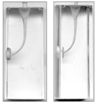
Figure 2 A scanned image of a gutta percha cone was inserted into shaped canals.
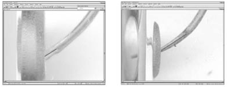
Figure 3 The distance from the apex of the prepared canal to the gutta percha cone tip was measured by an image analysis program (Image-Pro EXPRESS, MediaCybernetics, USA).
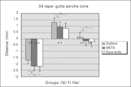
Figure 4 Distance from the apex of the prepared canal to the gutta percha cone tip according to the gutta percha cone brands. *indicates statistically significant difference between the brands of gutta percha cone (p < 0.05). PF .04: finally preparation with ProFile .04 #25 file. PT: finally preparation with ProTaper F2. HS .04: finally preparation with HEROShaper .04 #25 file.

Figure 5 Distance from the apex of the prepared canal to the tip of gutta percha cone according to the gutta percha cone brands. *indicates statistically significant difference between the brands of gutta percha cone (p < 0.05). PF .06: finally preparation with ProFile .06 #25 file. PT: finally preparation with ProTaper F2. HS .06: finally preparation with HEROShaper .06 #25 file.
Reference
-
1. Gutmann JL, Johnson WT. Cohen S, Hagreaves KM, editors. Obturation of the cleaned and shaped root canal system. Pathways of the pulp. 2006. 9th ed. St Louis: Mosby;358–399.2. Kratchman SI. Obturation of the root canal system. Dent Clin North Am. 2004. 48:203–215.
Article3. Hsu YY, Kim S. The ProFile system. Dent Clin North Am. 2004. 48:69–85.
Article4. Thomas C, Baumann MA. ProTaper NT system. Dent Clin North Am. 2004. 48:87–111.5. Veltri M, Mollo A. A comparative study of Endoflare-HeroShaper and Mtwo NiTi instruments in the preparation of curved root canal. Int Endod J. 2005. 38:610–616.6. Gordon MPJ, Love RM. An evaluation of .06 tapered gutta-percha cones for filling of .06 taper prepared curved root canals. Int Endod J. 2005. 38:87–96.
Article7. Bal AS, Hicks ML. Comparison of laterally condensed .06 and .02 tapered gutta-percha and sealer in vitro. J Endod. 2001. 27:786–788.
Article8. Hembrough MW, Steiman R. Lateral condensation in canals prepared with NiTi rotary instruments: An evaluation of the use of three different master cone. J Endod. 2002. 28:516–519.9. Calberson FLG, Deroose CAJG, Hommez GMG, Raes H, De Moor RJG. Shaping ability of GTTM rotary files in simulated resin root canals. Int Endod J. 2002. 35:607–614.
Article10. Eldeeb ME, Boraas JC. The effect of different files on the preparation shape of severely curved canals. Int Endod J. 1985. 18:1–7.
Article11. Kum KY, Spängberg L, Cha BY, Jung IY, Lee SJ, Lee CY. Shaping ability of three ProFile rotary instrumentation techniques in simulated resin root canals. J Endod. 2000. 26:719–723.
Article12. Dummer PMH, Alodeh MHA. A method for the construction of simulated root canals in clear resin blocks. Int Endod J. 1991. 24:63–66.
Article13. Thompson SA, Dummer PMH. Shaping ability of ProFile .04 taper series 29 rotary nickel-titanium instruments in simulated root canals. Part 1. Int Endod J. 1997. 30:1–7.14. Kazemi R, Stennman E, Spängberg LS. Machining efficiency and wear resistance of nickel-titanium endodontic files. Oral Surg Oral Med Oral Pathol Oral Radiol Endod. 1996. 81:596–602.
Article15. Yang GB, Zhou XD. Shaping ability of progressive versus constant taper instrumentations in simulated root canals. Int Endod J. 2006. 39:791–799.
Article16. Bergmans L, Cleynenbreugel JV. Progressive versus constant tapered shaft design using NiTi rotary instruments. Int Endod J. 2003. 36:288–295.
Article17. Kwon OS, Kim SK. Apical Fitness of Non-standardized Gutta-percha Cones in Simulated Root Canals Prepared with Rotary Root Canal Instruments. J Korean Acad Conserv Dent. 2000. 25:390–398.
- Full Text Links
-
- Actions
-
Cited
- CITED
-
- Close
- Share
-
- Similar articles
-
- A comparative study of the canal configuration after shaping by protaper rotary and hand files in resin simulated canals
- A study of insertion depth of buchanan plugger after shaping using NI-TI rotary files in simulated resin root canals
- The Effect of canal obturation according to the depth of the System B Plugger tip in the Type IV canal
- Obturation efficiency of non-standardized gutta-percha cone in curved root canals prepared with 0.06 taper nickel-titanium instruments
- Relative efficacy of three Ni-Ti file systems used by undergraduates
