Spinal Extradural Meningioma: A Case Report and Review of the Literature
- Affiliations
-
- 1Department of Radiology, Hallym University Sacred Heart Hospital, Hallym University College of Medicine, Anyang, Korea. silwater007@hallym.or.kr
- 2Department of Pathology, Hallym University Sacred Heart Hospital, Hallym University College of Medicine, Anyang, Korea.
- KMID: 2416400
- DOI: http://doi.org/10.3348/jksr.2018.79.1.11
Abstract
- Spinal meningiomas account for 12% of all the meningiomas and are usually located in the intradural extramedullary space. In some cases, they are associated with some extradural extensions. However, purely extradural spinal meningiomas are rare. Additionally, it is difficult to make an accurate preoperative diagnosis. We report a case of pathologically confirmed atypical meningioma, presented as a posterior epidural mass on the thoracic spine. We review the case, clinical symptoms, radiologic findings and the histologic features.
Figure
-
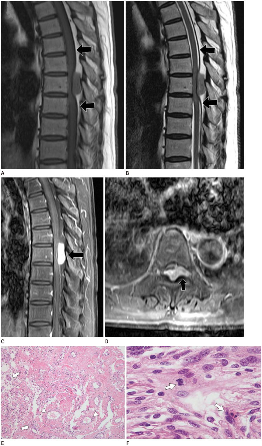
Fig. 1 MRI and pathologic findings of spinal extradual meningioma in a 58-year-old woman who has progressive weakness of both lower legs for 4 months. A, B. The T1-weighted image (A) and the T2-weighted image (B) display the posterior epidural mass with a similar signal intensity to the spinal cord. The subarachnoid space is obliterated at the level of the mass and the spinal cord is compressed. The dura mater is seen as a dark line (arrows) separating the extradural mass from the intradural structures. Elevated signal change, within the compressed spinal cord, represents compressive myelopathy on the T2-weighted image (B). C, D. Contrast-enhanced sagittal (C) and axial (D) T1-weighted images depict strong and homogeneous contrast enhancement of the posterior epidural mass (arrows). Note the complete filling of the posterior epidural space and the extension to the left neural foramina of the vertebrae (D). E. Histologically, the tumor was dominated by multiple vessels (arrowheads) interspersed with small meningothelial tumor cells (arrows) and focal necrosis (hematoxylin and eosin, × 100). F. Mitotic figures (arrows) are frequently identified, despite bland cytologic features of the tumor cells (hematoxylin and eosin, × 400).
Reference
-
1. Haranhalli N, Nakhla JP, Yassari R, Kinon MD. Radiographic pearls in the evaluation of an extradural thoracic meningioma: a case report. Cureus. 2017; 9:e1031.
Article2. Ben Nsir A, Boughamoura M, Mahmoudi H, Kilani M, Hattab N. Uncommon progression of an extradural spinal meningioma. Case Rep Surg. 2014; 2014:630876.
Article3. Plank C, Koller A, Mueller-Mang C, Bammer R, Thurnher MM. Diffusion-weighted MR imaging (DWI) in the evaluation of epidural spinal lesions. Neuroradiology. 2007; 49:977–985.
Article4. Frank BL, Harrop JS, Hanna A, Ratliff J. Cervical extradural meningioma: case report and literature review. J Spinal Cord Med. 2008; 31:302–305.
Article5. Jeong SK, Seong HY, Roh SW. Extra-intradural spinal meningioma: a case report. Korean J Spine. 2014; 11:202–204.
Article6. Yang T, Wu L, Yang C, Xu Y. Epidural angiomatous meningioma of the thoracic spine: a case report. Oncol Lett. 2016; 11:458–460.
Article7. Bettaswamy G, Ambesh P, Das KK, Sahu R, Srivastava A, Mehrotra A, et al. Extradural spinal meningioma: revisiting a rare entity. J Craniovertebr Junction Spine. 2016; 7:65–68.8. Khayal HB, Abograra A, Iashhab M. Extradural spinal meningioma in a nine-year-old girl: a case report and review of the literature. Open Access J Neurol Neurosurg. 2017; 5:555659.
Article9. El Khamary SM, Alorainy IA. Case 100: spinal epidural meningioma. Radiology. 2006; 241:614–617.
Article10. Klekamp J, Samii M. Surgical results for spinal meningiomas. Surg Neurol. 1999; 52:552–562.
Article
- Full Text Links
-
- Actions
-
Cited
- CITED
-
- Close
- Share
-
- Similar articles
-
- Extradural Spinal Lymphoplasmacyte-Rich Meningioma in the Thoracic Spine: A Case Report and Literature Review
- A Case of Thoracic Extradural Chordoid Meningioma: Focusing on Radiologic Features
- An Extradural En-Plaque Meningioma Involving Thoracic Spine
- Purely Extradural Spinal Meningioma of the Cervical Spine
- Extra-intradural Spinal Meningioma: A Case Report
