MRI Findings of Intercostal Schwannoma: A Case Report
- Affiliations
-
- 1Department of Radiology, Kangnam Sacred Heart Hospital, Hallym University College of Medicine, Seoul, Korea. younglady@hallym.or.kr
- 2Department of Pathology, Kangnam Sacred Heart Hospital, Hallym University College of Medicine, Seoul, Korea.
- KMID: 2416390
- DOI: http://doi.org/10.3348/jksr.2018.79.2.63
Abstract
- Intercostal schwannomas are uncommon, encapsulated neoplasms that originate in nerve sheaths of intercostal nerves. They account for less than 10% of primary neural tumors of the chest wall. Herein, we report a pathologically confirmed case of intercostal schwannoma with typical magnetic resonance imaging findings.
Figure
-
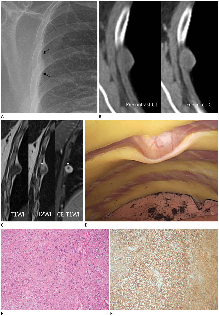
Fig. 1 A 61-year-old woman with intercostal schwannoma involving the right 5th intercostal nerve. A. Chest radiography shows a nodular lesion abutting on right upper chest wall, just inferior to 5th rib (arrows). B. Pre-enhanced CT demonstrates a 1.5 cm nodule showing heterogeneous attenuation in the chest wall, just inferior to the 5th rib. After CE, the mass shows thin peripheral enhancement. C. The mass reveals low signal intensity on T1WI, intermediate signal intensity with internal high signal intensity focus (arrowhead) on T2WI. On gadolinium-enhanced fat-suppressed T1WI, it shows bright enhancement, with tiny non-enhancing area (arrowhead) corresponding to the high signal intensity focus on T2WI. D. Intraoperative thoracoscopic image demonstrates a well circumscribed mass abutting on inferior margin of the right 5th rib showing mass effect on the chest wall. CE = contrast enhancement, CT = computed tomography, T1WI = T1-weighted image, T2WI = T2-weighted image E. Photomicrograph shows an Antoni A zone of compact cellular architecture with nuclear palisading (Verocay bodies) and Antoni B zone of spindle or rounded cells within loose myxoid stroma (hematoxylin and eosin stain, × 100). F. Photomicrograph shows diffuse strong positive finding in S100 immunohistochemical stain (× 100).
Reference
-
1. Meyer C, Rodepeter F, Bartsch D, Kirschbaum A. Intercostal neurinoma: a rare cause of persistent thoracic pain. Thorac Cardiovasc Surg Rep. 2014; 3:48–50.
Article2. Tateishi U, Gladish GW, Kusumoto M, Hasegawa T, Yokoyama R, Tsuchiya R, et al. Chest wall tumors: radiologic findings and pathologic correlation: part 2. malignant tumors. Radiographics. 2003; 23:1491–1150.3. McClenathan JH, Bloom RJ. Peripheral tumors of the intercostal nerves. Ann Thorac Surg. 2004; 78:713–714.
Article4. Nam SJ, Kim S, Lim BJ, Yoon CS, Kim TH, Suh JS, et al. Imaging of primary chest wall tumors with radiologic-pathologic correlation. Radiographics. 2011; 31:749–770.
Article5. Pavlus JD, Carter BW, Tolley MD, Keung ES, Khorashadi L, Lichtenberger JP 3rd. Imaging of thoracic neurogenic tumors. AJR Am J Roentgenol. 2016; 207:552–561.
Article6. Hwang ST, Sung DJ, Sim KC, Han NY, Park BJ, Kim MJ, et al. Radiologic findings of renal schwannoma: a case report and literature review. J Korean Soc Radiol. 2018; 78:289–294.
Article7. Nakazono T, White CS, Yamasaki F, Yamaguchi K, Egashira R, Irie H, et al. MRI findings of mediastinal neurogenic tumors. AJR Am J Roentgenol. 2011; 197:W643–W652.
Article8. Lin J, Martel W. Cross-sectional imaging of peripheral nerve sheath tumors: characteristic signs on CT, MR imaging, and sonography. AJR Am J Roentgenol. 2001; 176:75–82.9. Takanashi Y, Urabe N. [Schwannoma of the chest wall showing a bead-like appearance;report of a case]. Kyobu Geka. 2013; 66:1027–1029.10. Kim KS, Ji SR, Kim HM, Kwon YJ, Hwang JH, Lee SY. Intercostal nerve schwannoma encountered during a rib-latissimus dorsi osteomyocutaneous flap operation. Arch Plast Surg. 2015; 42:800–802.
Article
- Full Text Links
-
- Actions
-
Cited
- CITED
-
- Close
- Share
-
- Similar articles
-
- Refractory intercostal neuralgia due to intercostal schwannoma: A case report
- Hypervascular Vestibular Schwannoma: A Case Report
- Benign Schwannoma Mimicking Metastatic Lesion on F-18 FDG PET/CT in Differentiated Thyroid Cancer
- Diagnostic Value of MRI in Schwannoma
- Spinal Intradural Schwannoma with Torsion: A Case Report
