Contrast-free (Zero-contrast) TAVR for Severe Aortic Stenosis in Patient with Chronic Kidney Disease
- Affiliations
-
- 1Division of Cardiology, Department of Internal Medicine, Severance Cardiovascular Hospital, Yonsei University College of Medicine, Seoul, Korea. MKHONG61@yuhs.ac
- KMID: 2416259
- DOI: http://doi.org/10.12997/jla.2018.7.1.62
Abstract
- Transcatheter aortic valve replacement (TAVR) or transcatheter aortic valve implantation (TAVI) for severe aortic stenosis (AS) is a minimally invasive interventional procedure that repairs a valve without removing the old, damaged valve. Instead, a replacement valve is wedged into the location of the native aortic valve. During TAVR, contrast is used for conventional aortic root angiography, positioning of the TAVR valve device, and assessing the peripheral vasculature. Therefore, contrast-induced acute kidney injury (AKI) is a major concern when performing TAVR and is associated with increased mortality in patients with impaired renal function. Although the exact mechanism of post-TAVR AKI is unknown and appears multifactorial, contrast medium has been reported as a major contributing factor. We report a case of zero-contrast TAVR for severe AS in a patient with chronic kidney disease (CKD). The procedure was successfully performed with only fluoroscopic and transesophageal echocardiography (TEE) guidance.
Keyword
MeSH Terms
Figure
-

Fig. 1 Transthoracic echocardiogram showed severe AS (AVA: 0.96 cm2 by continuity equation) with peak/mean pressure gradient (64/33 mmHg). AS; aortic stenosis, AVA; aortic valve area, BP; blood pressure.

Fig. 2 Annulus dimension and perimeter derived diameter by 3-dimentional echocardiogram.

Fig. 3 Location of catheter was identified by using biplane TEE (dotted circle indicated tip of catheter and asterisk indicated catheter). TEE; transesophageal echocardiography, PAT; patient, T; temperature.
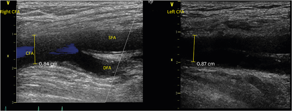
Fig. 4 Vascular ultrasound showed acceptable access site of left external iliac artery. CFA; common femoral artery, SFA; superficial femoral artery, DFA; deep femoral artery.
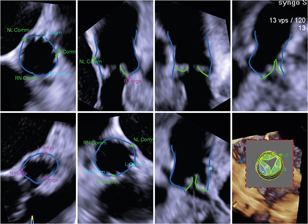
Fig. 5 3D-echo in pre-evaluation and TEE during TAVR. TEE; transesophageal echocardiography, TAVR; transcatheter aortic valve replacement, Comm; commissure.
Reference
-
1. Otto CM, Kumbhani DJ, Alexander KP, Calhoon JH, Desai MY, Kaul S, et al. 2017 ACC expert consensus decision pathway for transcatheter aortic valve replacement in the management of adults with aortic stenosis: a report of the American College of Cardiology Task Force on clinical expert consensus documents. J Am Coll Cardiol. 2017; 69:1313–1346.
Article2. Eskandari M, Aldalati O, Byrne J, Dworakowski R, Wendler O, Alcock E, et al. Zero contrast transfemoral transcatheter aortic valve replacement using fluoroscopy-echocardiography fusion imaging. Am J Cardiol. 2016; 117:1861–1862.
Article3. Cheungpasitporn W, Thongprayoon C, Kashani K. Transcatheter aortic valve replacement: a kidney's perspective. J Renal Inj Prev. 2016; 5:1–7.
Article4. Fassa AA, Himbert D, Vahanian A. Mechanisms and management of TAVR-related complications. Nat Rev Cardiol. 2013; 10:685–695.
Article5. Scherner M, Wahlers T. Acute kidney injury after transcatheter aortic valve implantation. J Thorac Dis. 2015; 7:1527–1535.6. Thongprayoon C, Cheungpasitporn W, Srivali N, Ungprasert P, Kittanamongkolchai W, Greason KL, et al. Acute kidney injury after transcatheter aortic valve replacement: a systematic review and meta-analysis. Am J Nephrol. 2015; 41:372–382.
Article7. Varga-Szemes A, Cannao PM, Muscogiuri G, De Cecco CN, Giri S, Piccini D, et al. Non-contrast 3D radial and QISS MRA for transcatheter aortic valve replacement planning. J Cardiovasc Magn Reson. 2015; 17:O71.
Article8. Voigtländer L, Schewel J, Martin J, Schewel D, Frerker C, Wohlmuth P, et al. Impact of kidney function on mortality after transcatheter valve implantation in patients with severe aortic valvular stenosis. Int J Cardiol. 2015; 178:275–281.
Article9. Bagur R, Webb JG, Nietlispach F, Dumont E, De Larochellière R, Doyle D, et al. Acute kidney injury following transcatheter aortic valve implantation: predictive factors, prognostic value, and comparison with surgical aortic valve replacement. Eur Heart J. 2010; 31:865–874.
Article10. Barbash IM, Ben-Dor I, Dvir D, Maluenda G, Xue Z, Torguson R, et al. Incidence and predictors of acute kidney injury after transcatheter aortic valve replacement. Am Heart J. 2012; 163:1031–1036.
Article11. Bagur R, Rodés-Cabau J, Doyle D, De Larochellière R, Villeneuve J, Lemieux J, et al. Usefulness of TEE as the primary imaging technique to guide transcatheter transapical aortic valve implantation. JACC Cardiovasc Imaging. 2011; 4:115–124.
Article
- Full Text Links
-
- Actions
-
Cited
- CITED
-
- Close
- Share
-
- Similar articles
-
- Transcatheter Aortic Valve Replacement with Minimal Contrast Dye in Patients with Renal Insufficiency
- Infective Endocarditis Associated with Transcatheter Aortic Valve Replacement: Potential Importance of Local Trauma for a Deadly Nidus
- Low Contrast and Low kV CTA Before Transcatheter Aortic Valve Replacement: A Systematic Review
- Aortic Stenosis: New Insights in Diagnosis, Treatment, and Prevention
- Spontaneous Contrast Echo on Two-Dimensional Echocardiography: Report of 4 Cases
