Hepatic Toxocariasis with Atypical CT and MR Imaging Findings: a Case Report
- Affiliations
-
- 1Department of Radiology, Chungnam National University Hospital, Chungnam National University School of Medicine, Daejeon, Korea. shinks@cnu.ac.kr
- KMID: 2415890
- DOI: http://doi.org/10.13104/imri.2018.22.2.113
Abstract
- Hepatic toxocariasis is a type of visceral larva migrans caused by the migration of second-stage larvae of certain nematodes such as Toxocara canis to the liver. Histologically, the condition is characterized by granulomatous lesions containing eosinophils and inflammatory cells. We report a case of hepatic toxocariasis with atypical clinical and radiologic findings presenting as distinct, solitary hepatic nodule detected in a middle-aged woman.
MeSH Terms
Figure
-
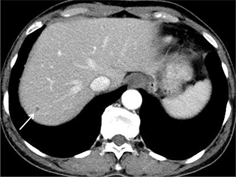
Fig. 1 Hepatic toxocariasis in a 59-year-old woman, CT findings. Contrast-enhanced abdominal CT at the level of the hepatic dome displays a tiny, relatively well-defined nodular lesion (arrow) with possible peripheral enhancement.

Fig. 2 Hepatic toxocariasis in a 59-year-old woman, MRI findings. (a) T1-weighted axial MR image shows a tiny, faintly hypointense nodule. (b) T2-weighted axial MR image shows a tiny, distinct nodule with bright signal intensity. (c) Diffusion-weighted image with a b value of 800 shows a hyperintense nodule. (d) The corresponding lesion is hypointense on the ADC map, representing diffusion restriction. (e-g) Gadolinium-enhanced T1-weighted images show a hypointense nodule with significant rim enhancement in the arterial phase (e) and with insignificant rim enhancement in the portal venous (f) and equilibrium (g) phases. (h) The hepatobiliary phase image shows hypointense nodule without rim enhancement. The arrow indicates the lesion in each image.
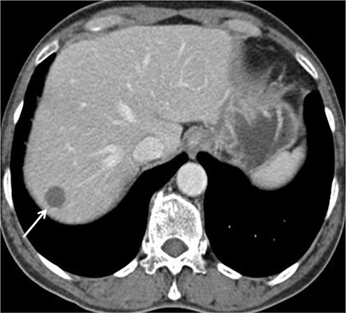
Fig. 3 Follow-up CT (6 months later). 2-phase abdominal CT image in the portal venous phase reveals a round, well-defined nodule (arrow) 1.5 cm in size with an attenuation of 31 Hounsfield unit.

Fig. 4 Hepatic toxocariasis in a 59-year-old woman, ultrasound findings. Ultrasound of the liver shows a round, slightly hypoechoic nodule (arrow) on the right hepatic dome.

Fig. 5 Hepatic toxocariasis in a 59-year-old woman, microscopic findings. Photomicrograph (Hematoxylin & Eosin, × 40) reveals hepatocyte necrosis (N) surrounded by a granulomatous inflammatory reaction consisting predominantly of eosinophils (arrowheads) and a multinucleated giant cell (arrow), suggestive of parasitic infection. The remnants of Toxocara canis larvae were not identified.

Fig. 6 Follow-up CT (2.5 years later). Two and a half years after treatment for Toxocara canis infection was begun, 3-phase abdominal dynamic CT image in the portal venous phase reveals a 1.0-cm lesion (arrow), which had decreased size from 1.5 cm at the beginning of treatment. The lesion's position has no changed, nor are there any new hepatic lesions.
Reference
-
1. Mukund A, Arora A, Patidar Y, et al. Eosinophilic abscesses: a new facet of hepatic visceral larva migrans. Abdom Imaging. 2013; 38:774–777.
Article2. Lim JH. Toxocariasis of the liver: visceral larva migrans. Abdom Imaging. 2008; 33:151–156.
Article3. Ishibashi H, Shimamura R, Hirata Y, Kudo J, Onizuka H. Hepatic granuloma in toxocaral infection: role of ultrasonography in hypereosinophilia. J Clin Ultrasound. 1992; 20:204–210.
Article4. Chang S, Lim JH, Choi D, et al. Hepatic visceral larva migrans of Toxocara canis: CT and sonographic findings. AJR Am J Roentgenol. 2006; 187:W622–W629.5. Dupas B, Barrier J, Barre P. Detection of Toxocara by computed tomography. Br J Radiol. 1986; 59:518–519.6. Arango CA. Visceral larva migrans and the hypereosinophilia syndrome. South Med J. 1998; 91:882–883.
Article7. Bhatia V, Batra Y, Acharya SK. Prophylactic phenytoin does not improve cerebral edema or survival in acute liver failure--a controlled clinical trial. J Hepatol. 2004; 41:89–96.
Article8. Hayashi K, Tahara H, Yamashita K, et al. Hepatic imaging studies on patients with visceral larva migrans due to probable Ascaris suum infection. Abdom Imaging. 1999; 24:465–469.
Article9. Laroia ST, Rastogi A, Sarin S. Case series of visceral larva migrans in the liver: CT and MRI findings. Int J Case Rep Imag. 2012; 3:7–12.
Article10. Azuma K, Yashiro N, Kinoshita T, Yoshigi J, Ihara N. Hepatic involvement of visceral larva migrans due to Toxocara canis: a case report--CT and MR findings. Radiat Med. 2002; 20:89–92.
- Full Text Links
-
- Actions
-
Cited
- CITED
-
- Close
- Share
-
- Similar articles
-
- Atypical Hepatic Angiomyolipoma: CT and MR Findings: A Case Report
- Pulmonary and Hepatic Involvement of Toxocariasis in an Adult: Case Report
- Liver Neoplasms: Atypical CT and MR Imaging Findings
- Hepatic Cavernous Hemangioma in Cirrhotic Liver: Imaging Findings
- Ultrasound, CT, and MR imaging Findings of Paraganglioma Originating at the Urinary Bladder: A Case Report
