MR Findings of Breast Implant Rupture Presenting with Unusual Breast Enlargement
- Affiliations
-
- 1Department of Radiology, Korea University Guro Hospital, Korea University College of Medicine, Seoul, Korea. wokhee@korea.ac.kr
- 2Department of Plastic Surgery, Korea University Guro Hospital, Korea University College of Medicine, Seoul, Korea.
- KMID: 2415889
- DOI: http://doi.org/10.13104/imri.2018.22.2.110
Abstract
- We report the case of a patient who presented with rupture of a silicone breast implant showing acute and chronic inflammation. Magnetic resonance imaging (MRI) showed silicone foci outside the implant shell and inside the pectoralis muscles that represented intra- and extracapsular ruptures of the implant and silicone granuloma. There were distinct fluid-fluid levels of various signal intensities and no signs of implant collapse such as "˜linguine sign.' Rather, we detected enlargement of both the implant shell and the breast.
MeSH Terms
Figure
-
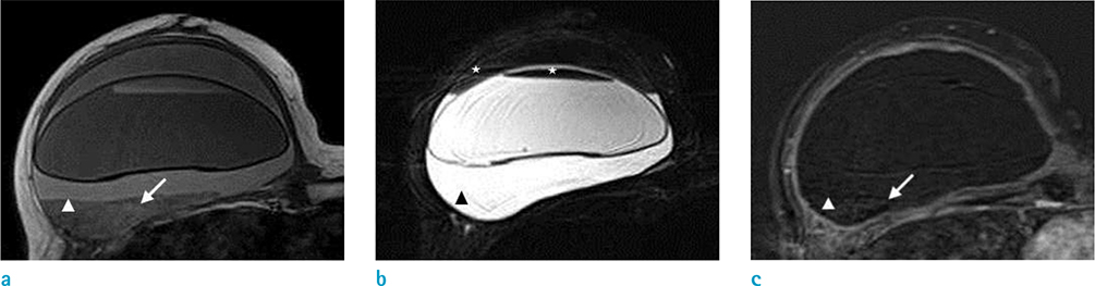
Fig. 1 T1- (a), fat-suppressed T2- (b), and 2 minutes delayed gadolinium-enhanced subtraction T1-weighted (c) MR images. A mass-like lesion of irregular shape and iso-signal intensity as compared with the silicone inside the implant shell in the posterior of the right breast represents silicone granuloma (arrowheads in a–c). It has small foci of subtle high signal intensity on T1WI with mild enhancement (arrows in a, c). Inside and outside the implant shell, a different fluid-fluid level and nondependent portion of low signal intensity on T2WI (asterisks in b) are noticed. Unlike other cases of silicone-implant rupture, this shows enlargement of breast and implant instead of the collapse usually expected.
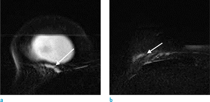
Fig. 2 Fat-suppressed T2-weighted MR images. Silicone gel of high signal intensity inside and outside the implant shell, and within pectoralis muscles superior and inferior to breast (arrows in a, b) represents intra- and extracapsular rupture.
Reference
-
1. Grubstein A, Cohen M, Steinmetz A, Cohen D. Siliconomas mimicking cancer. Clin Imaging. 2011; 35:228–231.
Article2. Yang N, Muradali D. The augmented breast: a pictorial review of the abnormal and unusual. AJR Am J Roentgenol. 2011; 196:W451–W460.
Article3. Colombo G, Ruvolo V, Stifanese R, Perillo M, Garlaschi A. Prosthetic breast implant rupture: imaging--pictorial essay. Aesthetic Plast Surg. 2011; 35:891–900.
Article4. Gorczyca DP, Gorczyca SM, Gorczyca KL. The diagnosis of silicone breast implant rupture. Plast Reconstr Surg. 2007; 120:49S–61S.
Article5. Berg WA, Nguyen TK, Middleton MS, Soo MS, Pennello G, Brown SL. MR imaging of extracapsular silicone from breast implants: diagnostic pitfalls. AJR Am J Roentgenol. 2002; 178:465–472.6. Orel SG. MR imaging of the breast. Radiol Clin North Am. 2000; 38:899–913.
Article7. Steinke K, Brook P, Ramuz O. Radiological pitfall: Siliconoma in internal mammary lymph node mimics breast cancer recurrence. Radiol Case Rep. 2011; 6:601.
Article8. Choi JJ, Lee JH, Kang BJ, et al. Clinical and imaging characteristics of Polyimplant Prosthesis hydrogel breast implants. J Comput Assist Tomogr. 2010; 34:449–455.
Article
- Full Text Links
-
- Actions
-
Cited
- CITED
-
- Close
- Share
-
- Similar articles
-
- Rupture of Breast Implants after Augmentation Mammoplasty: A Case Report of Simultaneous Intra-extracapsular Rupture
- Mechanical irritation by protruding bone: A possible cause of breast implant rupture
- Understanding Silicone Breast Implant-Associated Complications for Radiologists
- Complication of Augmentation Mammoplasty using Polysaccharide Hydrogel Breast implant: Two Cases Report
- The Reliability of Ultrasonographic Fending of Silicone Breast Implant Rupture
