Multi-vessel intractable coronary spasm development in a patient with aborted sudden cardiac death: a case study with intravascular ultrasound findings
- Affiliations
-
- 1Division of Cardiovascular Medicine, Department of Internal Medicine, Dankook University Hospital, Dankook University College of Medicine, Cheonan, Korea. neosoo70@dankook.ac.kr
- KMID: 2415745
- DOI: http://doi.org/10.12701/yujm.2018.35.1.121
Abstract
- Coronary spasm generally occurs in patients with minimal atherosclerotic plaque lesion, and it has a rather favorable prognosis. However, in some cases, coronary spasm may induce myocardial infarction and even sudden cardiac death (SCD). Here, we report a case in which multi-vessel intractable coronary vasospasm suddenly occurred in a diffuse atherosclerotic lesion after percutaneous coronary intervention (PCI) in a patient with aborted SCD. We identified the characteristics of the spasm portion in intravascular ultrasound (IVUS) images and conducted percutaneous cardiopulmonary bypass support-PCI with stenting as treatment. Intima and media thickening and a large attenuated plaque burden with rupture were identified in IVUS images at the obstructive spasm portion.
MeSH Terms
Figure
-

Fig. 1. Coronary angiography. (A) Initial left coronary angiography showing the significant stenosis of proximal LAD artery ostial lesion (white arrow). (B) Initial right coronary angiography showing the discrete significant stenosis of distal RCA (white arrow). *Coronary angiography after intracoronary nitrate injection. (C) Left coronary angiography after PCI and stenting at proximal LAD (white arrow). (D) Right coronary angiography after PCI and stenting at distal RCA (white arrow). LAD, left anterior descending; RCA, right coronary artery; PCI, percutaneous coronary intervention.

Fig. 2. Intravascular ultrasound. (A) Pre-intervention IVUS showing the eccentric echodense plaque with severe luminal narrowing at proximal LAD (white arrow), and there was not seen severe stenosis or plaque rupture at proximal LCx coronary artery (yellow arrow). (B) Post-intervention IVUS from distal LM to proximal LAD (white arrow: LAD and yellow arrow: LCx). LAD, left anterior descending; LCx, left circumflex; IVUS, intravascular ultrasound; PCI, percutaneous coronary intervention.

Fig. 3. Right coronary angiography and IVUS after spasm development. (A) Total occlusion of proximal RCA due to severe spasm (white arrow). (B) IVUS after resolved spasm after intracoronary nicorandil injection shows diffuse intima- and media-thickening with large amount of atheromatous plaque (yellow arrow) at mid RCA, and large attenuated plaque and prominent intima-thickening with rupture (white arrow) at proximal RCA. (C) PCI and stenting at proximal RCA (white arrow). RCA, right coronary artery; IVUS, intravascular ultrasound; PCI, percutaneous coronary intervention.

Fig. 4. Left coronary angiography after shock and recurrence AV block. (A) Total occlusion of proximal LCx artery due to severe spasm(white arrow). (B) Spasm resolved after intracoronary nicorandil injection (white arrow). AV, atrioventricular; LCx, left circumflex.
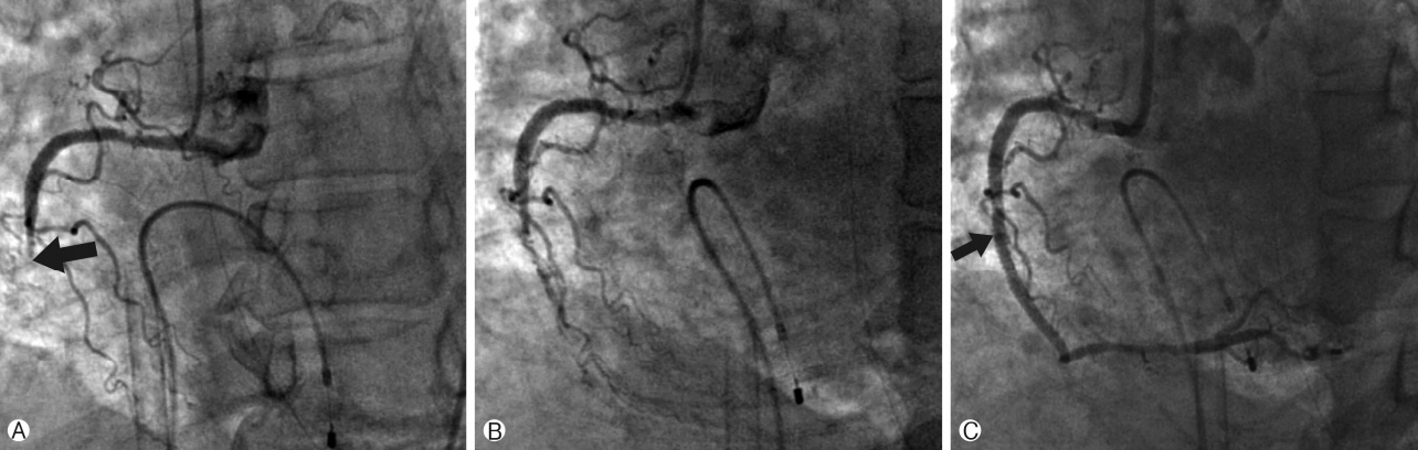
Fig. 5. Right coronary angiography after PCPS. (A) Total occlusion of mid RCA due to severe spasm (black arrow). (B) Spasm not fully resolved after several intracoronary nicorandil injections. (C) Mechanically breaking the spasm by stenting at mid RCA (black arrow). PCPS, percutaneous cardiopulmonary bypass support; RCA, right coronary artery.
Reference
-
1. JCS Joint Working Group. Guidelines for diagnosis and treatment of patients with vasospastic angina (Coronary Spastic Angina) (JCS 2013). Circ J. 2014; 78:2779–801.2. Yamagishi M, Miyatake K, Tamai J, Nakatani S, Koyama J, Nissen SE. Intravascular ultrasound detection of atherosclerosis at the site of focal vasospasm in angiographically normal or minimally narrowed coronary segments. J Am Coll Cardiol. 1994; 23:352–7.
Article3. Yasue H, Takizawa A, Nagao M, Nishida S, Horie M, Kubota J, et al. Long-term prognosis for patients with variant angina and influential factors. Circulation. 1988; 78:1–9.
Article4. Myerburg RJ, Kessler KM, Mallon SM, Cox MM, deMarchena E, Interian A Jr, et al. Life-threatening ventricular arrhythmias in patients with silent myocardial ischemia due to coronaryartery spasm. N Engl J Med. 1992; 326:1451–5.
Article5. Bhagwat A, Mukhedkar S. Severe generalized resistant spasm of the right coronary artery causing hemodynamic collapse after stenting. JACC Cardiovasc Interv. 2015; 8:e199–200.6. Miyao Y, Kugiyama K, Kawano H, Motoyama T, Ogawa H, Yoshimura M, et al. Diffuse intimal thickening of coronary arteries in patients with coronary spastic angina. J Am Coll Cardiol. 2000; 36:432–7.
Article7. Ota H, Kawase Y, Kondo H, Miyake T, Kamikawa S, Okubo M, et al. A case report of acute myocardial infarction induced by coronary spasm. Intravascular findings. Int Heart J. 2013; 54:237–9.8. Park YM, Kang WC, Shin KC, Han SH, Ahn T, Choi IS, et al. Repeated sudden cardiac death in coronary spasm: is IVUS helpful to decide treatment strategy? Int J Cardiol. 2012; 154:e57–9.
Article9. Shin ES, Ann SH, Singh GB, Lim KH, Yoon HJ, Hur SH, et al. OCT-defined morphological characteristics of coronary artery spasm sites in vasospastic angina. JACC Cardiovasc Imaging. 2015; 8:1059–67.
- Full Text Links
-
- Actions
-
Cited
- CITED
-
- Close
- Share
-
- Similar articles
-
- A Case of Aborted Sudden Cardiac Death during Exercise Associated with an Anomalous Origin of Right Coronary Artery
- A Case of Coronary Pseudostenosis, Diagnosed by Intravascular Ultrasound
- Repeated Aborted Sudden Cardiac Death with Long QT Syndrome in a Patient with Anomalous Origin of the Right Coronary Artery from the Left Coronary Cusp
- A case of three-vessel coronary artery spasm presenting with aborted sudden cardiac death
- Intravascular Ultrasonographic Detection of Atherosclerotic Lesions in Spastic Segments of the Coronary Arteries in Patients with Variant Angina
