J Clin Neurol.
2018 Jul;14(3):275-282. 10.3988/jcn.2018.14.3.275.
Age-Specific Cutoff Scores on a T1-Weighted Axial Medial Temporal-Lobe Atrophy Visual Rating Scale in Alzheimer's Disease Using Clinical Research Center for Dementia of South Korea Data
- Affiliations
-
- 1Department of Neurology, Ewha Womans University School of Medicine, Seoul, Korea. jjeong@ewha.ac.kr
- 2Department of Critical Care Medicine, Ewha Womans University School of Medicine, Seoul, Korea.
- 3Department of Preventive Medicine, Ewha Womans University School of Medicine, Seoul, Korea.
- 4Department of Neurology, Samsung Changwon Hospital, Sungkyunkwan University School of Medicine, Changwon, Korea.
- 5Department of Neurology, Konyang University Hospital, College of Medicine, Konyang University, Daejeon, Korea.
- 6Department of Neurology, Chonnam National University Medical School, Gwangju, Korea.
- 7Department of Neurology, College of Medicine, The Catholic University of Korea, Seoul, Korea.
- 8Department of Neurology, Sungkyunkwan University School of Medicine, Samsung Medical Center, Seoul, Korea.
- 9Department of Neurology, Pusan National University Hospital, Pusan National University School of Medicine and Medical Research Institute, Busan, Korea.
- 10Brain Fitness Center, Bobath Memorial Hospital, Seongnam, Korea.
- 11Department of Neurology, Myongji Hospital, Goyang, Korea.
- 12Department of Neurology, University of Ulsan College of Medicine, Asan Medical Center, Seoul, Korea.
- 13Department of Neurology, Dementia Center, Ilsan Hospital, National Health Insurance Service, Goyang, Korea.
- 14Department of Psychiatry, Pusan National University Hospital, Pusan National University School of Medicine and Medical Research Institute, Busan, Korea.
- 15Department of Neurology, Gachon University School of Medicine, Incheon, Korea.
- 16Department of Neurology, Dong-A University College of Medicine and Institute of Convergence Bio-Health, Busan, Korea.
- 17Department of Neurology, Seoul National University College of Medicine and Clinical Neuroscience Center of Seoul National University Bundang Hospital, Seongnam, Korea.
- 18Department of Neurology, Konkuk University Medical Center, Seoul, Korea.
- 19Department of Psychiatry, Asan Medical Center, University of Ulsan College of Medicine, Seoul, Korea.
- 20Department of Neurology, Eulji University College of Medicine, Daejeon, Korea.
- 21Department of Neurology, Ajou University School of Medicine, Suwon, Korea.
- 22Department of Neurology, Chung-Ang University College of Medicine, Seoul, Korea.
- 23Department of Neurology, Inha University School of Medicine, Incheon, Korea.
- KMID: 2415041
- DOI: http://doi.org/10.3988/jcn.2018.14.3.275
Abstract
- BACKGROUND AND PURPOSE
Visual assessment of medial temporal-lobe atrophy (MTA) has been quick, reliable, and easy to apply in routine clinical practice. However, one of the limitations in visual assessments of MTA is the lack of widely accepted age-adjusted norms and cutoff scores for MTA for a diagnosis of Alzheimer's disease (AD). This study aimed to determine the optimal cutoff score on a T1-weighted axial MTA Visual Rating Scale (VRS) for differentiating patients with AD from cognitively normal elderly people.
METHODS
The 3,430 recruited subjects comprising 1,427 with no cognitive impairment (NC) and 2003 AD patients were divided into age ranges of 50-59, 60-69, 70-79, and 80-89 years. Of these, 446 participants (218 in the NC group and 228 in the AD group) were chosen by random sampling for inclusion in this study. Each decade age group included 57 individuals, with the exception of 47 subjects being included in the 80- to 89-year NC group. The scores on the T1-weighted axial MTA VRS were graded by two neurologists. The cutoff values were evaluated from the area under the receiver operating characteristic curve.
RESULTS
The optimal axial MTA VRS cutoff score from discriminating AD from NC increased with age: it was ≥as ≥1, ≥2, and ≥3 in subjects aged 50-59, 60-69, 70-79, and 80-89 years, respectively (all p < 0.001).
CONCLUSIONS
These results show that the optimal cutoff score on the axial MTA VRS for diagnosing of AD differed according to the decade age group. This information could be of practical usefulness in the clinical setting.
Keyword
MeSH Terms
Figure
-

Fig. 1 A: Dimensions measured when scoring on the T1-weighted axial medial temporal-lobe atrophy VRS: width of the hippocampus (A′), width of the perimesencephalic cistern (C′), and width of the temporal horn (D′). B: Examples of grades 0 to 4 on the VRS. VRS: Visual Rating Scale.
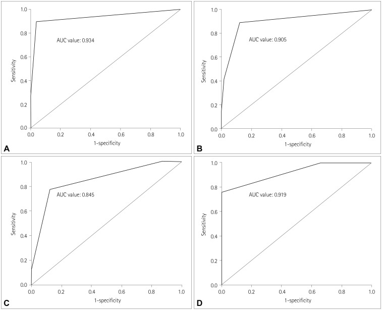
Fig. 2 AUC for optimal medial temporal-lobe atrophy Visual Rating Scale cutoff scores in subjects aged 50–59 years (A), 60–69 years (B), 70–79 years (C), and 80–89 years (D). AUC: area under the receiver operating characteristics curve.
Reference
-
1. Shen Q, Loewenstein DA, Potter E, Zhao W, Appel J, Greig MT, et al. Volumetric and visual rating of magnetic resonance imaging scans in the diagnosis of amnestic mild cognitive impairment and Alzheimer's disease. Alzheimers Dement. 2011; 7:e101–e108. PMID: 21784342.
Article2. Golde TE. The therapeutic importance of understanding mechanisms of neuronal cell death in neurodegenerative disease. Mol Neurodegener. 2009; 4:8. PMID: 19193222.
Article3. Jack CR Jr, Albert MS, Knopman DS, McKhann GM, Sperling RA, Carrillo MC, et al. Introduction to the recommendations from the National Institute on Aging-Alzheimer's Association workgroups on diagnostic guidelines for Alzheimer's disease. Alzheimers Dement. 2011; 7:257–262. PMID: 21514247.
Article4. Ferreira D, Cavallin L, Larsson EM, Muehlboeck JS, Mecocci P, Vellas B, et al. Practical cut-offs for visual rating scales of medial temporal, frontal and posterior atrophy in Alzheimer's disease and mild cognitive impairment. J Intern Med. 2015; 278:277–290. PMID: 25752192.
Article5. Duara R, Loewenstein DA, Potter E, Appel J, Greig MT, Urs R, et al. Medial temporal lobe atrophy on MRI scans and the diagnosis of Alzheimer disease. Neurology. 2008; 71:1986–1992. PMID: 19064880.
Article6. Harper L, Barkhof F, Fox NC, Schott JM. Using visual rating to diagnose dementia: a critical evaluation of MRI atrophy scales. J Neurol Neurosurg Psychiatry. 2015; 86:1225–1233. PMID: 25872513.
Article7. Kim GH, Kim JE, Choi KG, Lim SM, Lee JM, Na DL, et al. T1-weighted axial visual rating scale for an assessment of medial temporal atrophy in Alzheimer's disease. J Alzheimers Dis. 2014; 41:169–178. PMID: 24603942.
Article8. Scheltens P, Leys D, Barkhof F, Huglo D, Weinstein HC, Vermersch P, et al. Atrophy of medial temporal lobes on MRI in “probable” Alzheimer's disease and normal ageing: diagnostic value and neuropsychological correlates. J Neurol Neurosurg Psychiatry. 1992; 55:967–972. PMID: 1431963.
Article9. Visser PJ, Verhey FR, Hofman PA, Scheltens P, Jolles J. Medial temporal lobe atrophy predicts Alzheimer's disease in patients with minor cognitive impairment. J Neurol Neurosurg Psychiatry. 2002; 72:491–497. PMID: 11909909.10. Zhang Y, Qiu C, Lindberg O, Bronge L, Aspelin P, Bäckman L, et al. Acceleration of hippocampal atrophy in a non-demented elderly population: the SNAC-K study. Int Psychogeriatr. 2010; 22:14–25. PMID: 19958567.
Article11. Wu CC, Mungas D, Petkov CI, Eberling JL, Zrelak PA, Buonocore MH, et al. Brain structure and cognition in a community sample of elderly Latinos. Neurology. 2002; 59:383–391. PMID: 12177372.
Article12. Pereira JB, Cavallin L, Spulber G, Aguilar C, Mecocci P, Vellas B, et al. Influence of age, disease onset and ApoE4 on visual medial temporal lobe atrophy cut-offs. J Intern Med. 2014; 275:317–330. PMID: 24118559.
Article13. Choi SH, Kim S, Han SH, Na DL, Kim DK, Cheong HK, et al. Neurologic signs in relation to cognitive function in subcortical ischemic vascular dementia: a CREDOS (Clinical Research Center for Dementia of South Korea) study. Neurol Sci. 2012; 33:839–846. PMID: 22068220.
Article14. Barber R, Gholkar A, Scheltens P, Ballard C, McKeith IG, O'Brien JT. MRI volumetric correlates of white matter lesions in dementia with Lewy bodies and Alzheimer's disease. Int J Geriatr Psychiatry. 2000; 15:911–916. PMID: 11044873.
Article15. Ahn HJ, Chin J, Park A, Lee BH, Suh MK, Seo SW, et al. Seoul Neuropsychological Screening Battery-dementia version (SNSB-D): a useful tool for assessing and monitoring cognitive impairments in dementia patients. J Korean Med Sci. 2010; 25:1071–1076. PMID: 20592901.
Article16. Kim H, Na DL. Normative data on the Korean version of the Boston Naming Test. J Clin Exp Neuropsychol. 1999; 21:127–133. PMID: 10421007.17. Boutet C, Chupin M, Colliot O, Sarazin M, Mutlu G, Drier A, et al. Is radiological evaluation as good as computer-based volumetry to assess hippocampal atrophy in Alzheimer's disease? Neuroradiology. 2012; 54:1321–1330. PMID: 22782577.
Article18. Mossman D, Somoza E. ROC curves, test accuracy, and the description of diagnostic tests. J Neuropsychiatry Clin Neurosci. 1991; 3:330–333. PMID: 1821250.19. Claus JJ, Staekenborg SS, Holl DC, Roorda JJ, Schuur J, Koster P, et al. Practical use of visual medial temporal lobe atrophy cut-off scores in Alzheimer's disease: validation in a large memory clinic population. Eur Radiol. 2017; 27:3147–3155. PMID: 28083697.
Article20. Frisoni GB, Fox NC, Jack CR Jr, Scheltens P, Thompson PM. The clinical use of structural MRI in Alzheimer disease. Nat Rev Neurol. 2010; 6:67–77. PMID: 20139996.
Article21. Wollman DE, Prohovnik I. Sensitivity and specificity of neuroimaging for the diagnosis of Alzheimer's disease. Dialogues Clin Neurosci. 2003; 5:89–99. PMID: 22033599.22. Jonker C, Geerlings MI, Schmand B. Are memory complaints predictive for dementia? A review of clinical and population-based studies. Int J Geriatr Psychiatry. 2000; 15:983–991. PMID: 11113976.
Article23. Prestia A, Caroli A, Herholz K, Reiman E, Chen K, Jagust WJ, et al. Diagnostic accuracy of markers for prodromal Alzheimer's disease in independent clinical series. Alzheimers Dement. 2013; 9:677–686. PMID: 23375562.
Article24. Golebiowski M, Barcikowska M, Pfeffer A. Magnetic resonance imaging-based hippocampal volumetry in patients with dementia of the Alzheimer type. Dement Geriatr Cogn Disord. 1999; 10:284–288. PMID: 10364646.
Article25. Juottonen K, Laakso MP, Partanen K, Soininen H. Comparative MR analysis of the entorhinal cortex and hippocampus in diagnosing Alzheimer disease. AJNR Am J Neuroradiol. 1999; 20:139–144. PMID: 9974069.26. Raz N, Rodrigue KM, Head D, Kennedy KM, Acker JD. Differential aging of the medial temporal lobe: a study of a five-year change. Neurology. 2004; 62:433–438. PMID: 14872026.
Article27. Jack CR Jr, Petersen RC, Xu Y, O'Brien PC, Smith GE, Ivnik RJ, et al. Rate of medial temporal lobe atrophy in typical aging and Alzheimer's disease. Neurology. 1998; 51:993–999. PMID: 9781519.
Article28. Westman E, Cavallin L, Muehlboeck JS, Zhang Y, Mecocci P, Vellas B, et al. Sensitivity and specificity of medial temporal lobe visual ratings and multivariate regional MRI classification in Alzheimer's disease. PLoS One. 2011; 6:e22506. PMID: 21811624.
Article29. van der Flier WM, Pijnenburg YA, Fox NC, Scheltens P. Early-onset versus late-onset Alzheimer's disease: the case of the missing APOE ε4 allele. Lancet Neurol. 2011; 10:280–288. PMID: 21185234.
Article30. Fjell AM, Walhovd KB, Fennema-Notestine C, McEvoy LK, Hagler DJ, Holland D, et al. One-year brain atrophy evident in healthy aging. J Neurosci. 2009; 29:15223–15231. PMID: 19955375.
Article31. Xu J, Kobayashi S, Yamaguchi S, Iijima K, Okada K, Yamashita K. Gender effects on age-related changes in brain structure. AJNR Am J Neuroradiol. 2000; 21:112–118. PMID: 10669234.
- Full Text Links
-
- Actions
-
Cited
- CITED
-
- Close
- Share
-
- Similar articles
-
- Predictive Factors for Decline in Activities of Daily Living in Alzheimer's Disease Dementia with More than 2 Follow-up
- Association between Cognitive function, Behavioral and Psychological Symptoms of Dementia and Temporal Lobe Atrophy in Patients with Alzheimer's Disease and Mild Cognitive Impairment
- Impact of Vascular Risk Factors, Axial Medial Temporal Atrophy, White Matter Hyperintensity on Cognitive Outcome in Alzheimer's Diseases
- Posterior Type of Alzheimer's Dementia Presenting with Homonymous Hemianopsia
- Hippocampal Atrophy and Psychotic Symptoms in Patients with Alzheimer's Disease
