Guided bone regeneration using K-incision technique
- Affiliations
-
- 1Department of Periodontology, Seoul National University School of Dentistry, Seoul, Korea. guy@snu.ac.kr
- 2BK21 Plus Program, Dental Research Institute, Seoul National University, Seoul, Korea.
- KMID: 2414932
- DOI: http://doi.org/10.5051/jpis.2018.48.3.193
Abstract
- PURPOSE
The present study describes 3 patients with chronic periodontitis and consequent vertical resorption of the alveolar ridge who were treated using implant-based restoration with guided bone regeneration (GBR).
METHODS
After extraction of a periodontally compromised tooth, vertical bone augmentation using a K-incision was performed at the healed, low-level alveolar ridge.
RESULTS
The partial-split K-incision enabled soft tissue elongation without any change in buccal vestibular depth, and provided sufficient keratinized gingival tissue during GBR.
CONCLUSIONS
Within the limits of this study, the present case series demonstrated that the novel K-incision technique was effective for GBR and allowed normal implant-based restoration and maintenance of a healthy periodontal condition. However, further long-term follow-up and a large-scale randomized clinical investigation should be performed to evaluate the feasibility of this technique.
MeSH Terms
Figure
-
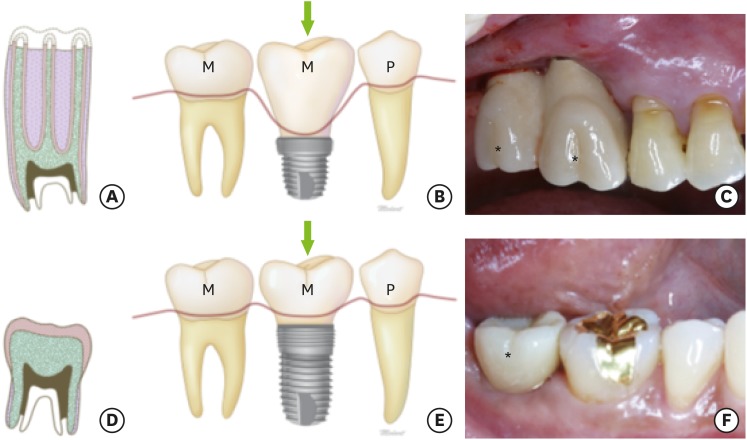
Figure 1 The terms ‘hypsodont’ and ‘brachydont.’ (A-C) ‘Hypsodont’ describes a tooth with large crowns. A schematic drawing of a hypsodont-like implant-based restoration is shown (marked with an arrow), as well as a clinical photo (marked with asterisks). (D-F) The term ‘brachydont’ refers to low-crowned teeth. A schematic drawing of a brachydont-like implant-based restoration is shown (marked with an arrow), as well as a clinical photo (marked with asterisks).M: molar, P: premolar.
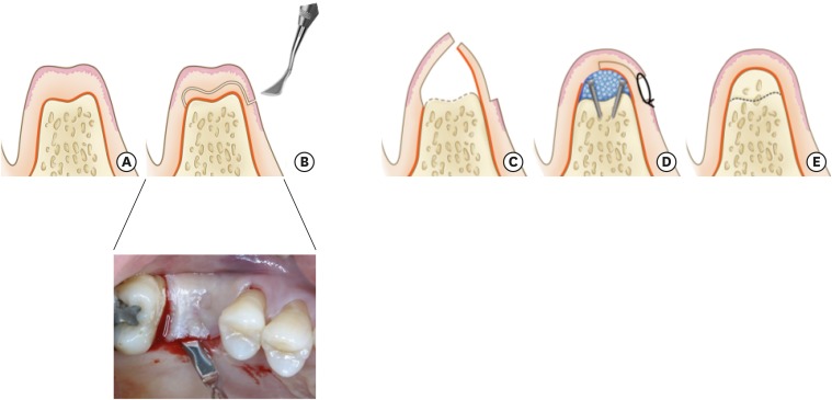
Figure 2 Schematic illustration of the K-incision. (A) Alveolar ridge showing a vertical bone defect. (B) A K-incision is performed using a Kirkland knife to split the gingiva into 2 parts. (C) After elevation of the gingival flap, decortication is performed to promote osteogenesis. (D) The microscrews are used as tent poles, and bovine bone mineral and membrane are applied. The divided gingival flap is then sutured. (E) After a 4-month healing period, the alveolar ridge shows vertically recovered bone with sufficient keratinized gingival tissue and an unchanged mucogingival junction level; the ridge is ready to receive an implant.
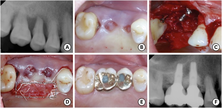
Figure 3 Case 1. (A) A periapical radiograph showing alveolar bone resorption around the teeth (#2 and #3). (B) An occlusal view of the tooth extraction site (teeth #2 and #3) after a 2-month healing period. (C) The microscrews were used as tent poles during guided bone regeneration. (D) The soft tissue extended by the K-incision is marked with a black tetragon. (E, F) A clinical photo (E) and radiograph (F) show the stable implant restoration with sufficient keratinized gingival tissue and alveolar bone over a 5-year periodic-recall follow-up period; no specific complications were observed.
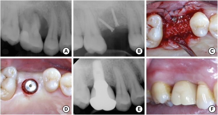
Figure 4 Case 2. (A) A periapical radiograph showing alveolar bone resorption around tooth #3. (B) Guided bone regeneration with microscrews in the vertically resorbed alveolar ridge. (C) Flap re-opening to remove the microscrews and to place the implant. (D) Sufficient keratinized gingiva around the implant. (E) A radiograph and (F) clinical photo showing the stable implant-based restoration over the 3-year periodic-recall follow-up period.
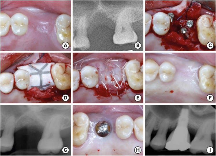
Figure 5 Case 3. (A) Occlusal view and (B) radiograph showing alveolar bone resorption around tooth #14 after a 3-month healing period. (C) The microscrews were used as tent poles, (D) inorganic bovine bone mineral particles and a titanium-reinforced membrane were applied, and (E) the flap was sutured with no tension. (F, G) The surgical site healed well, with proper alveolar bone width and height. (H) The dental implant was placed in the correct position. (I) A radiograph showing stable implant-based restoration over a 2-year periodic-recall follow-up.
Reference
-
1. Satloff D. Dental implant success. J Am Dent Assoc. 2002; 133:1040. 1042.
Article2. Zarb GA, Albrektsson T. Consensus report: towards optimized treatment outcomes for dental implants. J Prosthet Dent. 1998; 80:641. PMID: 9830066.3. Adell R, Lekholm U, Rockler B, Brånemark PI. A 15-year study of osseointegrated implants in the treatment of the edentulous jaw. Int J Oral Surg. 1981; 10:387–416. PMID: 6809663.
Article4. Jivraj S, Chee W. Treatment planning of implants in posterior quadrants. Br Dent J. 2006; 201:13–23. PMID: 16829878.
Article5. Buser D, Dula K, Belser UC, Hirt HP, Berthold H. Localized ridge augmentation using guided bone regeneration. II. surgical procedure in the mandible. Int J Periodontics Restorative Dent. 1995; 15:10–29. PMID: 7591520.6. Handelsman M. Surgical guidelines for dental implant placement. Br Dent J. 2006; 201:139–152. PMID: 16902543.
Article7. Garaicoa-Pazmiño C, Suárez-López del Amo F, Monje A, Catena A, Ortega-Oller I, Galindo-Moreno P, et al. Influence of crown/implant ratio on marginal bone loss: a systematic review. J Periodontol. 2014; 85:1214–1221. PMID: 24444399.
Article8. Birdi H, Schulte J, Kovacs A, Weed M, Chuang SK. Crown-to-implant ratios of short-length implants. J Oral Implantol. 2010; 36:425–433. PMID: 20545533.
Article9. Glantz PO, Nilner K. Biomechanical aspects of prosthetic implant-borne reconstructions. Periodontol 2000. 1998; 17:119–124. PMID: 10337319.
Article10. Lee KJ, Kim YG, Park JW, Lee JM, Suh JY. Influence of crown-to-implant ratio on periimplant marginal bone loss in the posterior region: a five-year retrospective study. J Periodontal Implant Sci. 2012; 42:231–236. PMID: 23346467.
Article11. Sanz I, Garcia-Gargallo M, Herrera D, Martin C, Figuero E, Sanz M. Surgical protocols for early implant placement in post-extraction sockets: a systematic review. Clin Oral Implants Res. 2012; 23(Suppl 5):67–79.
Article12. Vignoletti F, Matesanz P, Rodrigo D, Figuero E, Martin C, Sanz M. Surgical protocols for ridge preservation after tooth extraction. A systematic review. Clin Oral Implants Res. 2012; 23(Suppl 5):22–38.
Article13. Greenstein G, Greenstein B, Cavallaro J, Tarnow D. The role of bone decortication in enhancing the results of guided bone regeneration: a literature review. J Periodontol. 2009; 80:175–189. PMID: 19186957.
Article14. Nobre AM, Maló PS, Oliveira SH. The influence of implant location and position characteristics on peri-implant pathology. Eur J Prosthodont Restor Dent. 2014; 22:125–129. PMID: 25831714.15. Renvert S, Persson GR. Periodontitis as a potential risk factor for peri-implantitis. J Clin Periodontol. 2009; 36(Suppl 10):9–14. PMID: 19432626.
Article16. Warrer K, Buser D, Lang NP, Karring T. Plaque-induced peri-implantitis in the presence or absence of keratinized mucosa. An experimental study in monkeys. Clin Oral Implants Res. 1995; 6:131–138. PMID: 7578788.
Article17. Souza AB, Tormena M, Matarazzo F, Araújo MG. The influence of peri-implant keratinized mucosa on brushing discomfort and peri-implant tissue health. Clin Oral Implants Res. 2016; 27:650–655. PMID: 26474541.
Article18. Halperin-Sternfeld M, Zigdon-Giladi H, Machtei EE. The association between shallow vestibular depth and peri-implant parameters: a retrospective 6 years longitudinal study. J Clin Periodontol. 2016; 43:305–310. PMID: 26718112.19. Romanos GE. Periosteal releasing incision for successful coverage of augmented sites. A technical note. J Oral Implantol. 2010; 36:25–30. PMID: 20218867.
Article
- Full Text Links
-
- Actions
-
Cited
- CITED
-
- Close
- Share
-
- Similar articles
-
- Membrane Fixation using Healing Abutment during Guided Bone Regeneration: A Report of Two Cases
- Horizontal ridge augmentation using guided bone regeneration technique in two young patients with localized alveolar ridge defect: A case report
- The Sausage Technique using Collagen Membrane without Autogenous Bone Graft: A Case Report
- Titanium Mesh for Bone Augmentation in Oral Implant Surgery
- The Effect On Guided Bone Regeneration Of The Chitosan Membrane
