Ann Dermatol.
2018 Apr;30(2):226-228. 10.5021/ad.2018.30.2.226.
Recurrent Dermatofibrosarcoma Protuberans of Scalp in a Distant Location 10 Years after Primary Excision
- Affiliations
-
- 1Department of Dermatology, Korea University Ansan Hospital, Ansan, Korea. kumcihk@korea.ac.kr
- KMID: 2414687
- DOI: http://doi.org/10.5021/ad.2018.30.2.226
Abstract
- Dermatofibrosarcoma protuberans (DFSP) is a slow growing low-grade cutaneous sarcoma. Local recurrence after excision is common due to the poorly defined periphery that renders histological control of surgical margin difficult, Mohs micrographic surgery is the optimal method for treatment. A 41 years old male patient, who had a previous history of DFSP, came to our dermatology clinic for evaluation of an asymptomatic firm flesh-colored nodule on the forehead. Total excision biopsy was done and the mass was histologically proved as DFSP. Wide excision with reconstruction was performed and showed no sign of recurrence till 18-month follow up. Local recurrence is known to be common for DFSP but a new visible lesion distant from the initial site may be confused as a de novo lesion or a benign neoplasm especially in scalp area, and thus interrupt early detection of DFSP. Herein, we report a case of recurrent DFSP of scalp which recurred distant from the original lesion.
Keyword
MeSH Terms
Figure
-

Fig. 1 An asymptomatic firm flesh-colored nodule on the forehead.
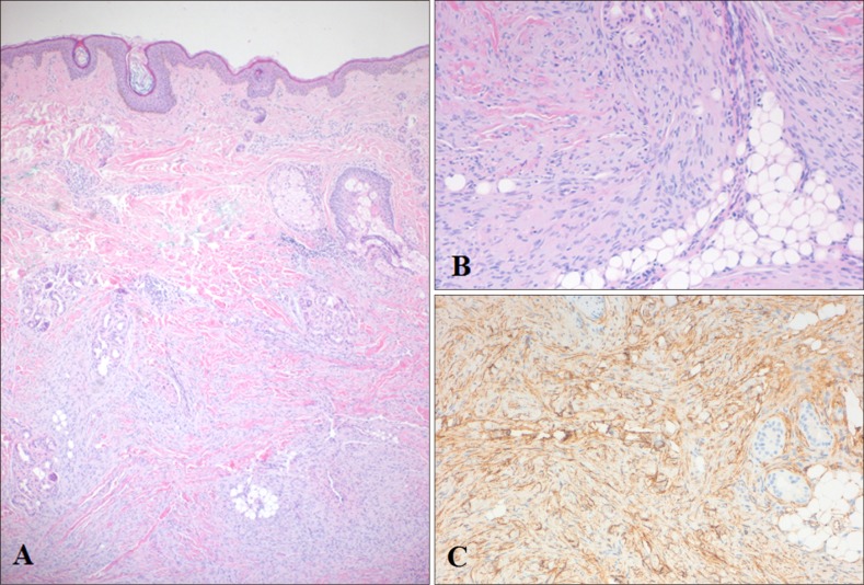
Fig. 2 (A) Densely packed spindle cells in storiform pattern (H&E, ×40). (B) Spindle cells with elongated nuclei infiltrates to fat layer and shows honeycomb appearance (H&E, ×100). (C) Immunohistochemistry results shows positivity for CD34 (CD34, ×100).
Reference
-
1. Kim MJ, Hur MS, Choi BG, Kim SY, Lee YW, Choe YB, et al. Pedunculated nodules as a variant of dermatofibrosarcoma protuberans. Ann Dermatol. 2016; 28:629–631. PMID: 27746644.
Article2. Ratner D, Thomas CO, Johnson TM, Sondak VK, Hamilton TA, Nelson BR, et al. Mohs micrographic surgery for the treatment of dermatofibrosarcoma protuberans. Results of a multiinstitutional series with an analysis of the extent of microscopic spread. J Am Acad Dermatol. 1997; 37:600–613. PMID: 9344201.3. Snow SN, Gordon EM, Larson PO, Bagheri MM, Bentz ML, Sable DB. Dermatofibrosarcoma protuberans: a report on 29 patients treated by Mohs micrographic surgery with long-term follow-up and review of the literature. Cancer. 2004; 101:28–38. PMID: 15221986.
Article4. Elgart GW, Hanly A, Busso M, Spencer JM. Bednar tumor (pigmented dermatofibrosarcoma protuberans) occurring in a site of prior immunization: immunochemical findings and therapy. J Am Acad Dermatol. 1999; 40:315–317. PMID: 10025857.
Article5. Harati K, Lange K, Goertz O, Lahmer A, Kapalschinski N, Stricker I, et al. A single-institutional review of 68 patients with dermatofibrosarcoma protuberans: wide re-excision after inadequate previous surgery results in a high rate of local control. World J Surg Oncol. 2017; 15:5. PMID: 28056985.
Article6. Thway K, Noujaim J, Jones RL, Fisher C. Dermatofibrosarcoma protuberans: pathology, genetics, and potential therapeutic strategies. Ann Diagn Pathol. 2016; 25:64–71. PMID: 27806849.
Article7. Gloster HM Jr, Harris KR, Roenigk RK. A comparison between Mohs micrographic surgery and wide surgical excision for the treatment of dermatofibrosarcoma protuberans. J Am Acad Dermatol. 1996; 35:82–87. PMID: 8682970.
- Full Text Links
-
- Actions
-
Cited
- CITED
-
- Close
- Share
-
- Similar articles
-
- A Case of Recurrent Dermatofibrosarcoma of the Scalp
- A Case of Recurrent Dermatofibrosarcoma Protuberans Treated by Mohs Micrographic Surgery Using Rush Permanent Sections
- Multiple Dermatofibrosarcoma Protuberans on the Scalp Treated by Tissue Expansion and Mohs Micrographic Surgery
- Two Cases of Recurrent Dermatofibrosarcoma Protuberans Treated by Mohs Micrographic Surgery
- Comments to “Pigmented Dermatofibrosarcoma Protuberans Presenting as a Faint Blue Macule in a Middle-aged Korean Womanâ€
