Breast Imaging Reporting and Data System Category 3 Lesions Detected on Whole-Breast Screening Ultrasound
- Affiliations
-
- 1Department of Radiology, Gachon University Gil Medical Center, Gachon University of Medicine and Science, Incheon, Korea.
- 2Department of Radiology and Center for Imaging Science, Samsung Medical Center, Sungkyunkwan University School of Medicine, Seoul, Korea. claudel@skku.edu
- KMID: 2413955
- DOI: http://doi.org/10.4048/jbc.2016.19.3.301
Abstract
- PURPOSE
This study assessed the incidence and cancer rate of probably benign lesions detected on bilateral whole-breast screening ultrasound (US), which corresponded to US Breast Imaging Reporting and Data System (BI-RADS) category 3, and evaluated the proper management of those lesions.
METHODS
This study was approved by the Institutional Review Board in our institution, which waived informed patient consent. We retrospectively reviewed US images of 1,666 patients who underwent bilateral whole-breast screening US as a supplemental screening test to negative screening mammography or screening US only. The incidence, clinical course, and cancer rate of screening US-detected probably benign lesions corresponding to US BI-RADS category 3 were investigated, and the size and multiplicity of screening US-detected category 3 lesions were evaluated.
RESULTS
Probably benign lesions corresponding to US BI-RADS category 3 were detected in 689 of 1,666 patients (41.4%) who underwent screening US. Among them, 653 had follow-up US images for at least 24 months, and among these 653, 190 (29.1%) had multiple bilateral category 3 lesions. Moreover, 539 of 1,666 patients (32.4%) had lesions ≤1 cm in size and 114 of 1,666 (6.8%) had lesions >1 cm (median, 0.82 cm; range, 0.3-4.2 cm). Four of the 653 patients (0.6%) showed suspicious interval changes and were categorized into BI-RADS category 4. Biopsy analysis confirmed only one lesion as invasive ductal carcinoma at the 6-month follow-up; another lesion was an intraductal papilloma and the remaining two were fibroadenomas. Overall cancer rate of the screening US-detected BI-RADS category 3 lesions was 0.2%.
CONCLUSION
The incidence of category 3 lesions detected on screening US only was very high, but the cancer rate was very low. Therefore, in an average-risk population, routine screening US is preferable over short-term follow-up for BI-RADS category 3 lesions detected on whole-breast screening US.
Keyword
MeSH Terms
Figure
-

Figure 1 Flow chart of study population.US=ultrasound; BI-RADS=Breast Imaging Reporting and Data System; F/U=follow-up.
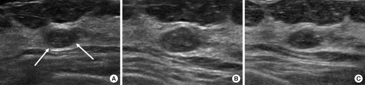
Figure 2 A 52-year-old woman with a mass detected on the screening breast ultrasound (US) in left breast, upper center. (A) An initial US image shows a 1.0 cm oval shape circumscribed isoechoic mass corresponding to Breast Imaging Reporting and Data System (BI-RADS) category 3 on US (arrows). (B) and (C) at the 12 months and 2 years follow-up US, the mass in the left upper center was not changed, and downgraded to BI-RADS category 2.
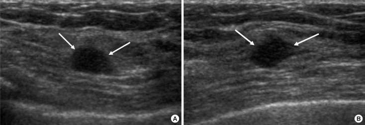
Figure 3 A 40-year-old woman with a nodule detected on the screening breast ultrasound (US) in right breast, upper inner quadrant. (A) An initial US image shows a 0.5 cm oval circumscribed isoechoic mass, corresponding to category 3 (arrows). (B) On the 6-month follow-up US, the surrounding tissue around the previous mass consisted of nodule together, and the nodule showed more district angular margin, more decreased echogenicity and increased size, and was assessed as category 4 (arrows). US-guided core needle biopsy revealed invasive ductal carcinoma, and the patient underwent right partial mastectomy.
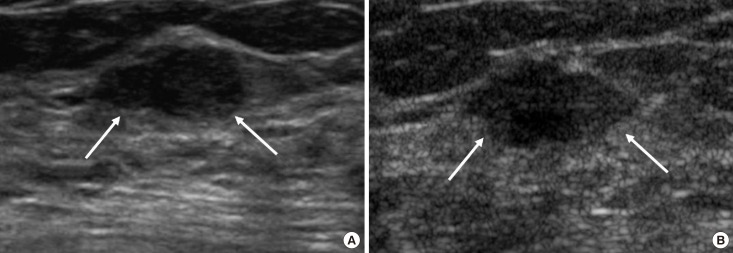
Figure 4 A 45-year-old woman with a mass detected on the screening breast ultrasound (US) in right breast, upper outer quadrant. (A) Initial US image showed a 1.2 cm oval circumscribed isoechoic mass in the right upper outer quadrant, corresponding to category 3 (arrows). (B) On the 12 month follow-up US, the margin of the mass changed into more microlobulated and indistinct, and was assessed as category 4 (arrows). US-guided core needle biopsy was performed and fibroadenoma was confirmed.
Reference
-
1. Kopans DB. The 2009 U.S. Preventive Services Task Force guidelines ignore important scientific evidence and should be revised or withdrawn. Radiology. 2010; 256:15–20. PMID: 20574081.
Article2. Buchberger W, DeKoekkoek-Doll P, Springer P, Obrist P, Dünser M. Incidental findings on sonography of the breast: clinical significance and diagnostic workup. AJR Am J Roentgenol. 1999; 173:921–927. PMID: 10511149.
Article3. Gordon PB, Goldenberg SL. Malignant breast masses detected only by ultrasound: a retrospective review. Cancer. 1995; 76:626–630. PMID: 8625156.
Article4. Kaplan SS. Clinical utility of bilateral whole-breast US in the evaluation of women with dense breast tissue. Radiology. 2001; 221:641–649. PMID: 11719658.
Article5. Kolb TM, Lichy J, Newhouse JH. Occult cancer in women with dense breasts: detection with screening US-diagnostic yield and tumor characteristics. Radiology. 1998; 207:191–199. PMID: 9530316.
Article6. Kolb TM, Lichy J, Newhouse JH. Comparison of the performance of screening mammography, physical examination, and breast US and evaluation of factors that influence them: an analysis of 27,825 patient evaluations. Radiology. 2002; 225:165–175. PMID: 12355001.
Article7. Moon WK, Noh DY, Im JG. Multifocal, multicentric, and contralateral breast cancers: bilateral whole-breast US in the preoperative evaluation of patients. Radiology. 2002; 224:569–576. PMID: 12147858.
Article8. Crystal P, Strano SD, Shcharynski S, Koretz MJ. Using sonography to screen women with mammographically dense breasts. AJR Am J Roentgenol. 2003; 181:177–182. PMID: 12818853.
Article9. Leconte I, Feger C, Galant C, Berlière M, Berg BV, D'Hoore W, et al. Mammography and subsequent whole-breast sonography of nonpalpable breast cancers: the importance of radiologic breast density. AJR Am J Roentgenol. 2003; 180:1675–1679. PMID: 12760942.10. Benson SR, Blue J, Judd K, Harman JE. Ultrasound is now better than mammography for the detection of invasive breast cancer. Am J Surg. 2004; 188:381–385. PMID: 15474430.
Article11. Berg WA, Gutierrez L, NessAiver MS, Carter WB, Bhargavan M, Lewis RS, et al. Diagnostic accuracy of mammography, clinical examination, US, and MR imaging in preoperative assessment of breast cancer. Radiology. 2004; 233:830–849. PMID: 15486214.
Article12. Berg WA, Blume JD, Cormack JB, Mendelson EB, Lehrer D, Böhm-Vélez M, et al. Combined screening with ultrasound and mammography vs mammography alone in women at elevated risk of breast cancer. JAMA. 2008; 299:2151–2163. PMID: 18477782.
Article13. Kelly KM, Dean J, Comulada WS, Lee SJ. Breast cancer detection using automated whole breast ultrasound and mammography in radiographically dense breasts. Eur Radiol. 2010; 20:734–742. PMID: 19727744.
Article14. Maskarinec G, Nagata C, Shimizu H, Kashiki Y. Comparison of mammographic densities and their determinants in women from Japan and Hawaii. Int J Cancer. 2002; 102:29–33. PMID: 12353230.
Article15. Shen YC, Chang CJ, Hsu C, Cheng CC, Chiu CF, Cheng AL. Significant difference in the trends of female breast cancer incidence between Taiwanese and Caucasian Americans: implications from age-period-cohort analysis. Cancer Epidemiol Biomarkers Prev. 2005; 14:1986–1990. PMID: 16103449.
Article16. Maskarinec G, Pagano I, Chen Z, Nagata C, Gram IT. Ethnic and geographic differences in mammographic density and their association with breast cancer incidence. Breast Cancer Res Treat. 2007; 104:47–56. PMID: 17009106.
Article17. Mendelson EB, Böhm-Vélez M, Berg WA, Whitman GJ, Feldman MI, Madjar H, et al. ACR BI-RADS ultrasound. In : D'Orsi CJ, Sickles EA, Mendelson EB, Morris EA, editors. ACR BI-RADS Atlas: Breast Imaging Reporting and Data System. Reston: American College of Radiology;2013.18. Kim SJ, Chang JM, Cho N, Chung SY, Han W, Moon WK. Outcome of breast lesions detected at screening ultrasonography. Eur J Radiol. 2012; 81:3229–3233. PMID: 22591758.
Article19. Barr RG, Zhang Z, Cormack JB, Mendelson EB, Berg WA. Probably benign lesions at screening breast US in a population with elevated risk: prevalence and rate of malignancy in the ACRIN 6666 trial. Radiology. 2013; 269:701–712. PMID: 23962417.
Article20. Chang JM, Koo HR, Moon WK. Radiologist-performed hand-held ultrasound screening at average risk of breast cancer: results from a single health screening center. Acta Radiol. 2015; 56:652–658. PMID: 24951614.
Article21. Moon HJ, Jung I, Park SJ, Kim MJ, Youk JH, Kim EK. Comparison of cancer yields and diagnostic performance of screening mammography vs. supplemental screening ultrasound in 4394 women with average risk for breast cancer. Ultraschall Med. 2015; 36:255–263. PMID: 24764212.22. Feig S. Cost-effectiveness of mammography, MRI, and ultrasonography for breast cancer screening. Radiol Clin North Am. 2010; 48:879–891. PMID: 20868891.
Article23. Irwig L, Houssami N, van Vliet C. New technologies in screening for breast cancer: a systematic review of their accuracy. Br J Cancer. 2004; 90:2118–2122. PMID: 15150556.
Article24. Moon HJ, Kim EK, Kwak JY, Yoon JH, Kim MJ. Interval growth of probably benign breast lesions on follow-up ultrasound: how can these be managed? Eur Radiol. 2011; 21:908–918. PMID: 21113596.
Article25. Stavros AT, Thickman D, Rapp CL, Dennis MA, Parker SH, Sisney GA. Solid breast nodules: use of sonography to distinguish between benign and malignant lesions. Radiology. 1995; 196:123–134. PMID: 7784555.
Article26. Skaane P, Engedal K. Analysis of sonographic features in the differentiation of fibroadenoma and invasive ductal carcinoma. AJR Am J Roentgenol. 1998; 170:109–114. PMID: 9423610.
Article27. Kim SJ, Ko EY, Shin JH, Kang SS, Mun SH, Han BK, et al. Application of sonographic BI-RADS to synchronous breast nodules detected in patients with breast cancer. AJR Am J Roentgenol. 2008; 191:653–658. PMID: 18716090.
Article28. Buchberger W, Niehoff A, Obrist P, DeKoekkoek-Doll P, Dünser M. Clinically and mammographically occult breast lesions: detection and classification with high-resolution sonography. Semin Ultrasound CT MR. 2000; 21:325–336. PMID: 11014255.
Article29. Corsetti V, Houssami N, Ferrari A, Ghirardi M, Bellarosa S, Angelini O, et al. Breast screening with ultrasound in women with mammography-negative dense breasts: evidence on incremental cancer detection and false positives, and associated cost. Eur J Cancer. 2008; 44:539–544. PMID: 18267357.
Article
- Full Text Links
-
- Actions
-
Cited
- CITED
-
- Close
- Share
-
- Similar articles
-
- Breast Imaging Reporting and Data System (BI-RADS): Advantages and Limitations
- Categorization and Evaluation of Usefulness of Breast Lesions with using Ultrasound BI-RADS (Breast Imaging Reporting and Data system)
- Usefulness of ultrasound elastography in reducing the number of Breast Imaging Reporting and Data System category 3 lesions on ultrasonography
- Thyroid Imaging Reporting and Data System (TIRADS)
- Validity of Breast Cancer Symptom Questionnaire and Its Relationship With Breast Ultrasonography in Young Female Night Workers
