Recurrent symptomatic cemento-osseous dysplasia: A case report
- Affiliations
-
- 1Department of Oral and Maxillofacial Radiology, School of Dentistry, Chonbuk National University, Jeonju, Korea. beam@jbnu.ac.kr
- KMID: 2413925
- DOI: http://doi.org/10.5624/isd.2018.48.2.131
Abstract
- Cemento-osseous dysplasia (COD) is a benign fibro-osseous lesion of bone, in which normal bone is replaced by fibrous tissue, followed by calcification with osseous and cementum-like tissue. COD is classified into 3 categories according to its location: periapical, focal, and florid COD (FCOD). On radiography, FCOD appears radiolucent in its early stages. As it matures, radiopacities appear within the lesion, causing them to show a mixed appearance of radiolucency and radiopacity. Because FCOD is usually asymptomatic and grows in a self-limited manner, it does not require treatment. Secondary infection is the most frequent cause of symptomatic cases. We report a case of FCOD with symptoms that appeared after a dental restoration procedure and persisted after repeated operations. The purpose of this report is to emphasize the importance of thorough radiological evaluations of patients with FCOD before treatment.
Keyword
MeSH Terms
Figure
-
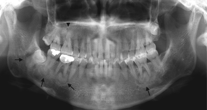
Fig. 1 Panoramic image shows multiple sclerotic masses with radiolucent rims in the apical region of the left mandibular second molar and the right mandibular first, second, and third molars (arrows), as well as completely radiopaque masses in the apical region of the right maxillary first molar and the left maxillary second premolar (arrowhead).
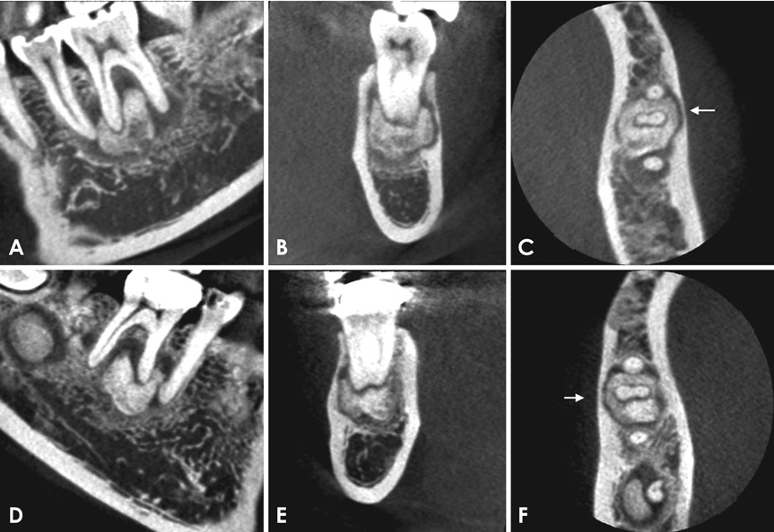
Fig. 2 Cone-beam computed tomography scans reveal well-defined radiopaque masses surrounded by radiolucent rims in the periapical regions of the left mandibular second premolar and first molar (A–C) and the right mandibular first and second molar (D–F). All the lesions were separated from the adjacent teeth by a radiolucent line. Invasion of the cortical bone can be observed (arrows). (A and D: sagittal, B and E: frontal, C and F: axial scan)

Fig. 3 Scintigraphs show increased radiopharmaceutical uptake in the posterior maxilla and mandible.
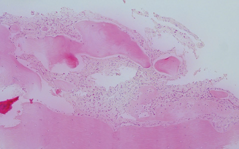
Fig. 4 Histopathologic examination shows multiple areas of irregular woven bone in dense fibrous stroma without any fibrous capsules, representing cemento-osseous dysplasia (H&E stain, original magnification ×100).

Fig. 5 Panoramic image 3 years after surgery shows the remaining calcified masses around the right mandibular first molar and increased periapical radiolucency at the right mandibular second molar.
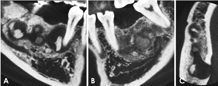
Fig. 6 A and B. Cone-beam computed tomography (CBCT) scans 3 years after surgery reveal the remaining and newly-formed radiopacities inside the lesion. C. Axial CBCT scan shows perforation of the lingual cortical plate near the right mandibular second molar.
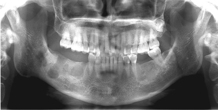
Fig. 7 Panoramic image obtained immediately after secondary surgery shows that all the lesions of the right mandible had been removed.

Fig. 8 Histopathologic finding shows numerous areas of woven bone in fibrous connective tissue with infiltration of inflammatory cells (H&E stain, original magnification ×40).

Fig. 9 Panoramic image demonstrates sclerotic masses surrounded by radiolucent halos in the right and left mandible at the previous surgical sites.
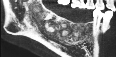
Fig. 10 Sagittal cone-beam computed tomography scan clearly reveals sclerotic masses surrounded by radiolucent halos in the right mandible at the previous surgical sites.
Cited by 1 articles
-
Cemento-osseous dysplasia: clinical presentation and symptoms
Inhye Nam, Jihye Ryu, Sang-Hun Shin, Yong-Deok Kim, Jae-Yeol Lee
J Korean Assoc Oral Maxillofac Surg. 2022;48(2):79-84. doi: 10.5125/jkaoms.2022.48.2.79.
Reference
-
1. MacDonald DS. Maxillofacial fibro-osseous lesions. Clin Radiol. 2015; 70:25–36.
Article2. MacDonald-Jankowski DS. Florid cemento-osseous dysplasia: a systematic review. Dentomaxillofac Radiol. 2003; 32:141–149.
Article3. Mainville GN, Turgeon DP, Kauzman A. Diagnosis and management of benign fibro-osseous lesions of the jaws: a current review for the dental clinician. Oral Dis. 2017; 23:440–450.
Article4. Su L, Weathers DR, Waldron CA. Distinguishing features of focal cemento-osseous dysplasia and cemento-ossifying fibromas. II. A clinical and radiologic spectrum of 316 cases. Oral Surg Oral Med Oral Pathol Oral Radiol Endod. 1997; 84:540–549.5. Cavalcanti PH, Nascimento EH, Pontual ML, Pontual AD, Marcelos PG, Perez DE, et al. Cemento-osseous dysplasias: imaging features based on cone beam computed tomography scans. Braz Dent J. 2018; 29:99–104.
Article6. El-Mofty SK, Nelson B, Toyosawa S, Wright JM. Cementoosseous dysplasia. In : El-Naggar AK, Chan JK, Rubin Grandis J, Takata T, Slootweg PJ, editors. International Agency for Research on Cancer. WHO classification of head and neck tumours. 4th ed. Lyon: International Agency for Research on Cancer;2017. p. 254–255.7. MacDonald-Jankowski DS. Fibro-osseous lesions of the face and jaws. Clin Radiol. 2004; 59:11–25.
Article8. Mortazavi H, Baharvand M, Rahmani S, Jafari S, Parvaei P. Radiolucent rim as a possible diagnostic aid for differentiating jaw lesions. Imaging Sci Dent. 2015; 45:253–261.
Article9. Singer SR, Mupparapu M, Rinaggio J. Florid cemento-osseous dysplasia and chronic diffuse osteomyelitis Report of a simultaneous presentation and review of the literature. J Am Dent Assoc. 2005; 136:927–931.10. Cavalcante MB, de Oliveira Lima AL, Júnior MA, Santos MB. Florid cemento-osseous dysplasia simultaneous the chronic suppurative osteomyelitis in mandible. J Craniofac Surg. 2016; 27:2173–2176.
Article11. Delai D, Bernardi A, Felippe GS, da Silveira Teixeira C, Felippe WT, Santos Felippe MC. Florid cemento-osseous dysplasia: a case of misdiagnosis. J Endod. 2015; 41:1923–1926.
Article12. Dağistan S, Tozoğlu Ü, Göregen M, Çakur B. Florid cemento-osseous dysplasia: a case report. Med Oral Patol Oral Cir Bucal. 2007; 12:E348–E350.13. Wakasa T, Kawai N, Aiga H, Kishi K. Management of florid cemento-osseous dysplasia of the mandible producing solitary bone cyst: report of a case. J Oral Maxillofac Surg. 2002; 60:832–835.
Article14. Bencharit S, Schardt-Sacco D, Zuniga JR, Minsley GE. Surgical and prosthodontic rehabilitation for a patient with aggressive florid cemento-osseous dysplasia: a clinical report. J Prosthet Dent. 2003; 90:220–224.
Article15. Toffanin A, Benetti R, Manconi R. Familial florid cemento-osseous dysplasia: a case report. J Oral Maxillofac Surg. 2000; 58:1440–1446.
Article16. Önder B, Kurşun Ş, Öztaş B, Barş E, Erdem E. Florid osseous dysplasia in a middle-aged Turkish woman: a case report. Imaging Sci Dent. 2013; 43:197–200.
Article17. Kawai T, Hiranuma H, Kishino M, Jikko A, Sakuda M. Cemento-osseous dysplasia of the jaws in 54 Japanese patients: a radiographic study. Oral Surg Oral Med Oral Pathol Oral Radiol Endod. 1999; 87:107–114.18. Esfahanizadeh N, Yousefi H. Successful implant placement in a case of florid cemento-osseous dysplasia: a case report and literature review. J Oral Implantol. (in press).
Article19. Kim JH, Song BC, Kim SH, Park YS. Clinical, radiographic, and histological findings of florid cemento-osseous dysplasia: a case report. Imaging Sci Dent. 2011; 41:139–142.
Article20. Resnick CM, Novelline RA. Cemento-osseous dysplasia, a radiological mimic of periapical dental abscess. Emerg Radiol. 2008; 15:367–374.
Article21. Huh JK, Shin SJ. Misdiagnosis of florid cemento-osseous dysplasia leading to unnecessary root canal treatment: a case report. Restor Dent Endod. 2013; 38:160–166.
Article22. Eskandarloo A, Yousefi F. CBCT findings of periapical cemento-osseous dysplasia: a case report. Imaging Sci Dent. 2013; 43:215–218.
Article23. Said-al-Naief NA, Surwillo E. Florid osseous dysplasia of the mandible: report of a case. Compend Contin Educ Dent. 1999; 20:1017–1019.24. Smith S, Patel K, Hoskinson AE. Periapical cemental dysplasia: a case of misdiagnosis. Br Dent J. 1998; 185:122–123.
Article25. Consolaro A. Florid cemento-osseous dysplasia: one of the few contraindications to osseointegrated implants. Dent Press Implantol. 2015; 9:26–33.
Article26. Macdonald-Jankowski DS. Focal cemento-osseous dysplasia: a systematic review. Dentomaxillofac Radiol. 2008; 37:350–360.
Article27. Petrocelli M, Kretschmer W. Conservative treatment and implant rehabilitation of the mandible in a case of craniofacial fibrous dysplasia: a case report. J Oral Maxillofac Surg. 2014; 72:902.e1–902.e6.
Article28. Gerlach RC, Dixon DR, Goksel T, Castle JT, Henry WA. Case presentation of florid cemento-osseous dysplasia with concomitant cemento-ossifying fibroma discovered during implant explantation. Oral Surg Oral Med Oral Pathol Oral Radiol. 2013; 115:e44–e52.
Article
- Full Text Links
-
- Actions
-
Cited
- CITED
-
- Close
- Share
-
- Similar articles
-
- 3 Types of Cemento-Osseous Dysplasia: Case Reports
- Cemento-Osseous Dysplasia Largely Occurring in the Mandible: Case Report
- Cemento-osseous dysplasia: clinical presentation and symptoms
- Florid cemento-osseous dysplasia: a report of two cases
- CBCT findings of periapical cemento-osseous dysplasia: A case report
