Calcaneal Apophyseal Avulsion Fractures with Achilles Tendon Rupture in a 10-Year-Old Patient: A Case Report
- Affiliations
-
- 1Department of Orthopaedic Surgery, School of Medicine, Chosun University, Gwangju, Korea. leejy88@chosun.ac.kr
- KMID: 2413716
- DOI: http://doi.org/10.14193/jkfas.2018.22.2.74
Abstract
- Calcaneal apophysitis is a relatively common disease in young athletes. On the other hand, if not treated properly, it can lead to apophyseal avulsion fracture in rare cases. In the case of apophyseal avulsion fractures, it is often necessary to remove or preserve the bone fragment, which often requires a suture of the Achilles tendon. A 10-year-old badminton athlete visited the outpatients' clinic with pain in both heels from 10 months ago without any trauma history. After conservative therapy, the pain in the left heel was relived but the right heel pain persisted. After 10 months of conservative therapy, the patient visited the outpatients' clinic showing a calcaneal apophyseal avulsion fracture with a total rupture of the Achilles tendon. In the operation room, a bone fragment needed to be removed because of its poor viability and the fragment was too thin for fixation. After removing the bone fragment, the ruptured Achilles tendon was fixed with an anchor system.
Figure
-

Figure 1. Representive T1-weighted (A) and T2-weighted (B) proton density sagittal magnetic resonance image illustrating complete rupture of Achilles tendon. Demonstrating avulsion fracture and Achilles tendon retraction.
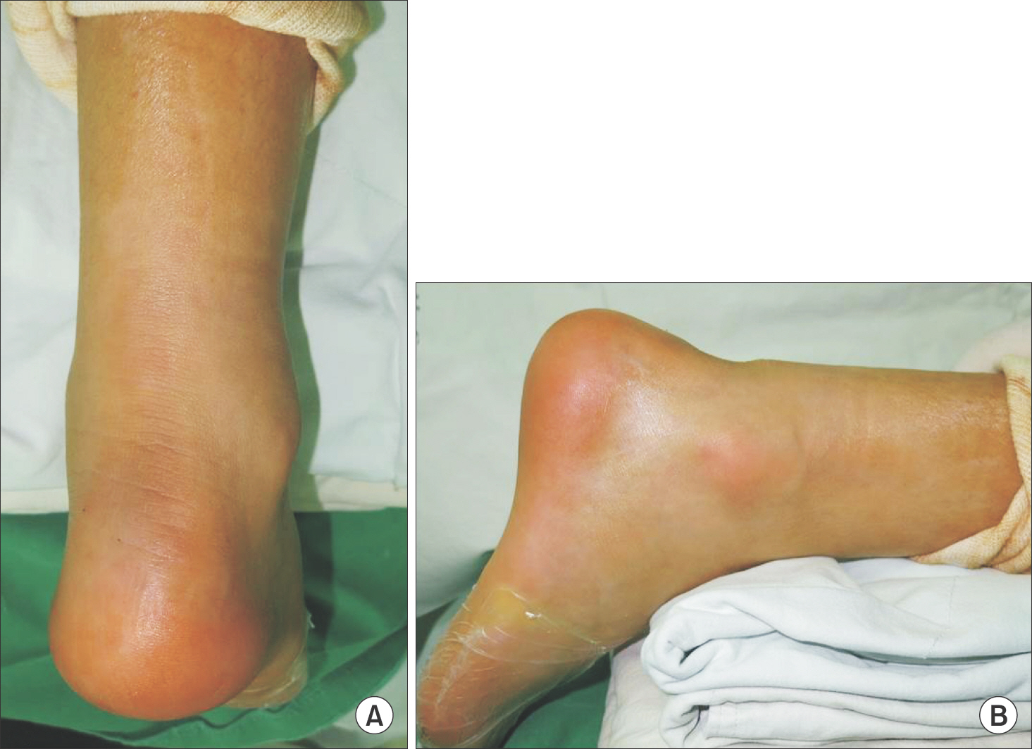
Figure 2. Skin dimpling of right heel. (A) Posterior aspct of ankle. (B) Lateral view.
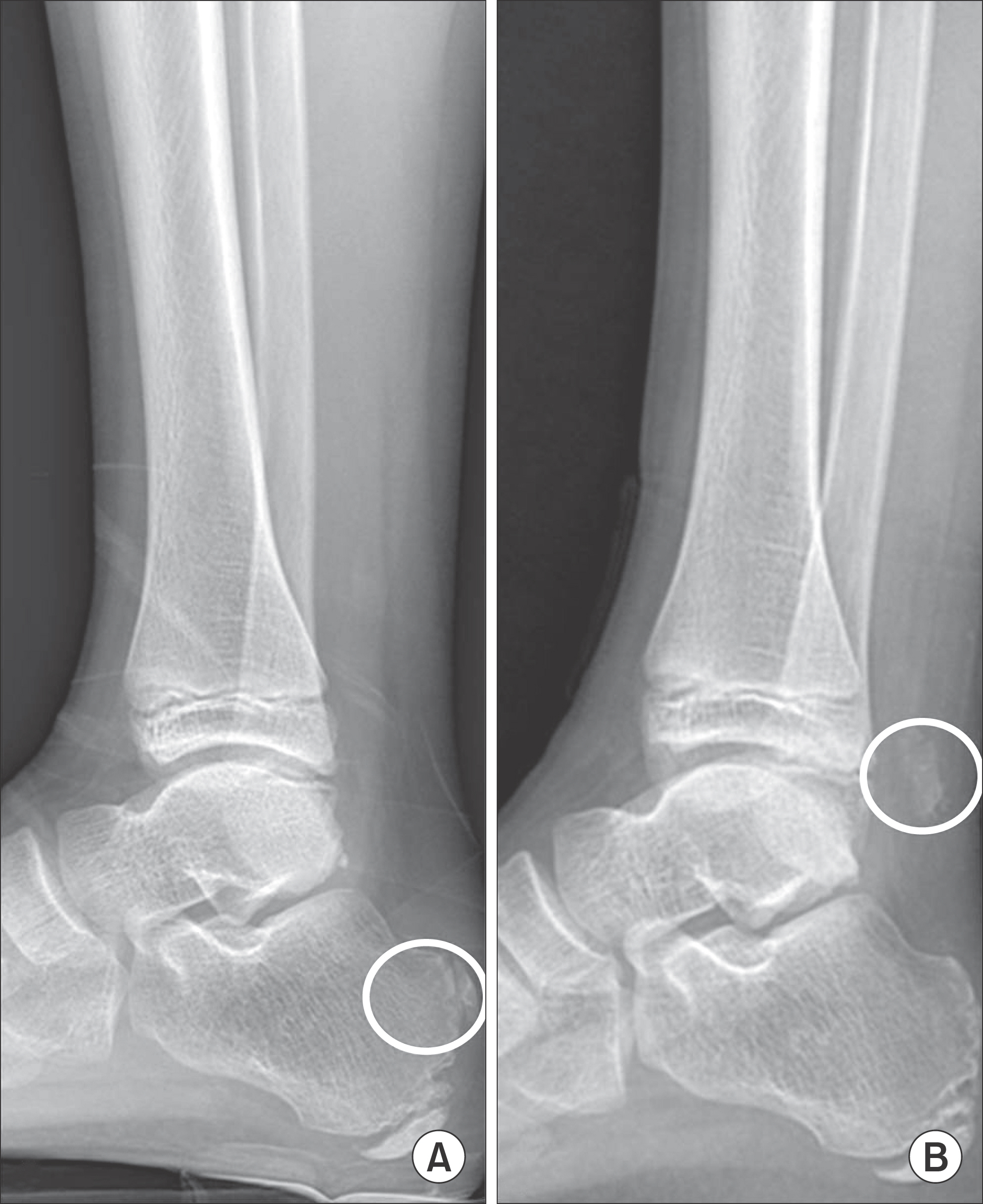
Figure 3. Bone fragment translation. (A) Initial simple radiograph. (B) Ten months later preoperative simple radiograph.
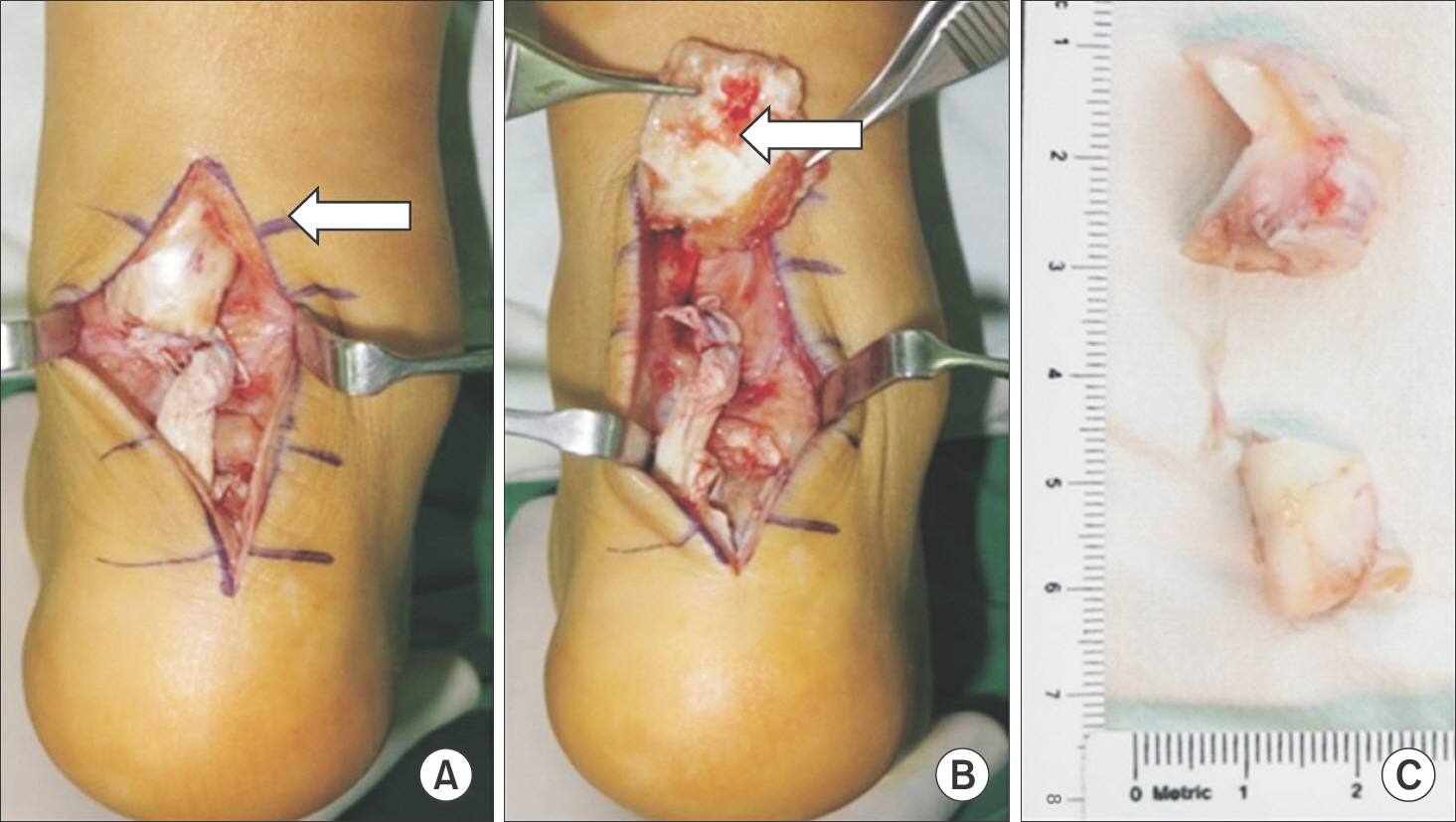
Figure 4. (A, B) Operative findings with bone fragment at the proximal segment of Achilles tendon (arrows). (C) Bone fragment.

Figure 5. Operative procedure with anchor system. (A) Insert anchor by pushing into calcaneal tuberosity. (B) Achilles repair by ankor suture.
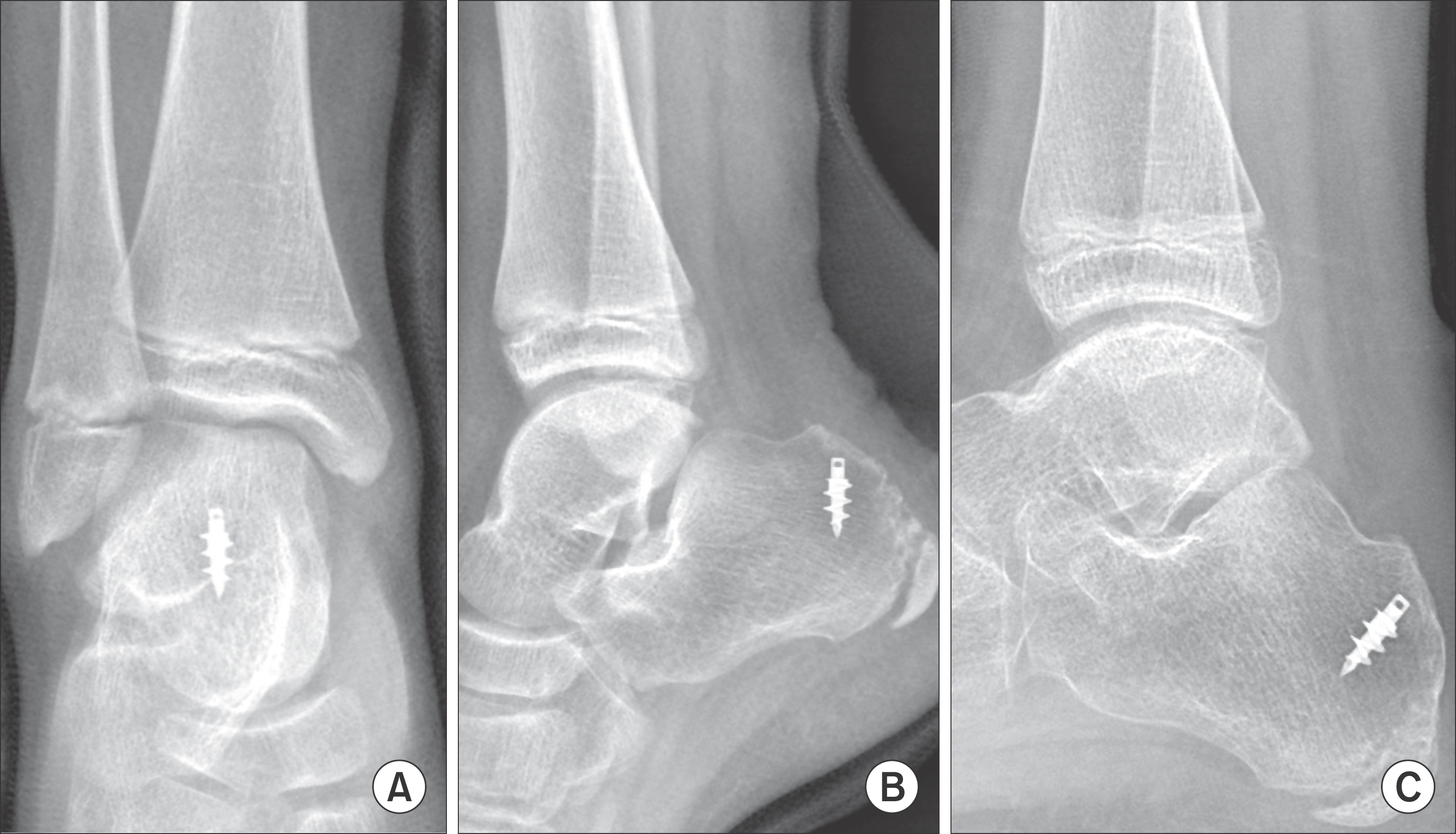
Figure 6. (A, B) Postoperative anteroposterior and lateral plain radiography. (C) Last follow-up, postoperative day 6 months lateral plain radiography.
Reference
-
1.Schmidt TL., Weiner DS. Calcaneal fractures in children. An evaluation of the nature of the injury in 56 children. Clin Orthop Relat Res. 1982. 171:150–5.
Article2.Mooney V. Avulsion of the epiphysis of the os calcis. J Bone Joint Surg. 1935. 17:1056–7.3.Fernandez JA., Vaquero MV., Cimiano JG. Epiphysiolysis of the great tuberosity of the calcaneum. J Bone Joint Surg Br. 1989. 71:321.4.Imai Y., Kitano T., Nakagawa K., Takaoka K. Calcaneal apophyseal avulsion fracture. Arch Orthop Trauma Surg. 2007. 127:331–3.
Article5.Lee KT., Young KW., Park YU., Park SY., Kim KC. Neglected Sever’s disease as a cause of calcaneal apophyseal avulsion fracture: case report. Foot Ankle Int. 2010. 31:725–8.
Article6.Cole RJ., Brown HP., Stein RE., Pearce RG. Avulsion fracture of the tuberosity of the calcaneus in children. A report of four cases and review of the literature. J Bone Joint Surg Am. 1995. 77:1568–71.
Article7.Paley KJ., Milem CA., Ebraheim NA., Merritt TR. Severe displacement of a calcaneal apophyseal fracture. J Foot Ankle Surg. 1994. 33:180–3.8.Ogden JA., Ganey TM., Hill JD., Jaakkola JI. Sever’s injury: a stress fracture of the immature calcaneal metaphysis. J Pediatr Orthop. 2004. 24:488–92.
Article
- Full Text Links
-
- Actions
-
Cited
- CITED
-
- Close
- Share
-
- Similar articles
-
- Calcaneal Tuberosity Avulsion Fracture after Repair of Achilles Tendon Rupture: A Case Report
- Achilles Tendon Sleeve Avulsion
- Operative Treatment of Avulsion Fractures of the Calcaneal Tuberosity
- Avulsion Fracture of Calcaneal Apophysis in an Adolescent Gymnast: A Case Report
- Achilles Tendon Rupture Associated With Ipsilateral Medial Malleolar Fracture (A Case Report)
