Is It Better to Enter a Volume CT Dose Index Value before or after Scan Range Adjustment for Radiation Dose Optimization of Pediatric Cardiothoracic CT with Tube Current Modulation?
- Affiliations
-
- 1Department of Radiology and Research Institute of Radiology, University of Ulsan College of Medicine, Asan Medical Center, Seoul 05505, Korea. ghw68@hanmail.net
- KMID: 2413698
- DOI: http://doi.org/10.3348/kjr.2018.19.4.692
Abstract
OBJECTIVE
To determine whether the body size-adapted volume computed tomography (CT) dose index (CTD(vol)) in pediatric cardiothoracic CT with tube current modulation is better to be entered before or after scan range adjustment for radiation dose optimization.
MATERIALS AND METHODS
In 83 patients, cardiothoracic CT with tube current modulation was performed with the body size-adapted CTDIvol entered after (group 1, n = 42) or before (group 2, n = 41) scan range adjustment. Patient-related, radiation dose, and image quality parameters were compared and correlated between the two groups.
RESULTS
The CTDIvol after the CT scan in group 1 was significantly higher than that in group 2 (1.7 ± 0.1 mGy vs. 1.4 ± 0.3 mGy; p < 0.0001). Image noise (4.6 ± 0.5 Hounsfield units [HU] vs. 4.5 ± 0.7 HU) and image quality (1.5 ± 0.6 vs. 1.5 ± 0.6) showed no significant differences between the two (p > 0.05). In both groups, all patient-related parameters, except body density, showed positive correlations (r = 0.49-0.94; p < 0.01) with the CTDIvol before and after the CT scan. The CTDIvol after CT scan showed modest positive correlation (r = 0.49; p ≤ 0.001) with image noise in group 1 but no significant correlation (p > 0.05) in group 2.
CONCLUSION
In pediatric cardiothoracic CT with tube current modulation, the CTDIvol entered before scan range adjustment provides a significant dose reduction (18%) with comparable image quality compared with that entered after scan range adjustment.
Keyword
Figure
-

Fig. 1 Axial CT image obtained approximately 1–2 cm above dome of liver in which X-ray output in CTDIvol based on 32-cm phantom was individually determined.To measure area and mean density, CT technologist draws region of interest to include entire patient cross-section with upper (50000 HU) and lower (−900 HU) limits of CT numbers.A. On same axial CT image with mediastinal window setting, AP was measured from most anterior body surface to most posterior body surface (vertical arrow) and LAT was measured from most right lateral body surface to most left lateral body surface (horizontal arrow). B. On same axial CT image with lung window setting, additional radiolucent pad (white arrow) is shown to be placed on CT table (black arrows) to adjust patient's vertical position at isocenter. Blanket wrapping around patient and patient cloth are shown in lung window. AP = anteroposterior diameter, CT = computed tomography, CTDIvol = volume CT dose index, HU = Hounsfield units, LAT = lateral diameter

Fig. 2 Comparison of user-determined radiation dose in pediatric cardiothoracic CT with tube current modulation in two groups.A. CT scout image shows slice position (horizontal orange line) of axial CT image for determination of CTDIvol that was entered after scan range adjustment (transparent purple rectangle) in group 1. B. CT scout image shows slice position (horizontal orange line) of axial CT image for determination of CTDIvol that was entered with minimal longitudinal range at same position (transparent purple rectangle) in group 2. C. In group 2, scan range was subsequently extended longitudinally (arrows) to full scan range (transparent purple rectangle) of cardiothoracic CT scanning. As result, initially entered CTDIvol value with minimal longitudinal range was automatically changed to new value after scan range adjustment, based on body attenuation information obtained from CT scout image in group 2. Of note, both arms are raised up on CT scout image in both cases.
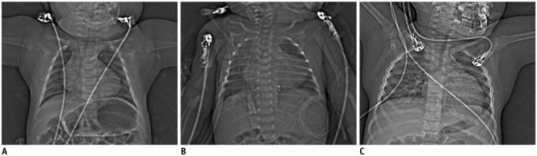
Fig. 3 Variable arm positions on CT scout images.A. CT scout image shows that both arms are horizontal, which is greatly disadvantageous in terms of image quality and radiation dose, as compared with both arms raised up. B. CT scout image shows that both arms are down beside body trunk, which may slightly degrade image quality and increase radiation dose, as compared with both arms raised up. C. CT scout image shows that right arm is raised up and left arm is horizontal, which may cause asymmetric image quality degradation in left shoulder region and increased radiation dose, as compared with both arms raised up.

Fig. 4 Subjective image quality grading of pediatric cardiothoracic CT.Axial (A) and coronal (B) CT images show excellent image quality (grade 1) without substantial artifacts in 6 months-old female infant whose arm were raised up during CT scanning. Axial CT image (A) at same slice position, where CTDIvol value was calculated, was used to measure CT densities in aorta (1), spinal muscles (2), and air (3) by placing rectangular regions of interest in areas showing homogeneous attenuation as much as possible. Axial (C) and coronal (D) CT images show severely degrade subjective image quality (grade 3) in posterior thoracic inlet and left shoulder regions due to horizontal position of left arm during CT scanning. Metal artifacts from electrocardiography electrode located at right upper chest are also noted on axial CT image (C).
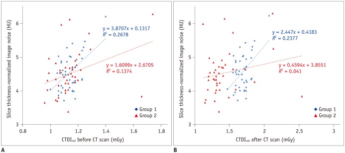
Fig. 5 Scatter plots demonstrating correlations between radiation dose and image noise of cardiothoracic CT.A. In scatter plot, group 1 illustrates higher correlation (r = 0.52; p = 0.0004) between CTDIvol before CT scan and slice thickness-normalized image noise than that (r = 0.37; p = 0.02) in group 2. B. In scatter plot, group 1 illustrates modest correlation (r = 0.49; p = 0.001) between CTDIvol after CT scan and slice thickness-normalized image noise. In contrast, group 2 shows no significant correlation (r = 0.20; p = 0.2) between them that may be attributed to reduction of CTDIvol after CT scan, graphically recognized as leftward shift of red triangular points of group 2 in X-axis, compared with blue rhomboid points of group 1.
Cited by 5 articles
-
Semiautomatic Three-Dimensional Threshold-Based Cardiac Computed Tomography Ventricular Volumetry in Repaired Tetralogy of Fallot: Comparison with Cardiac Magnetic Resonance Imaging
Hyun Woo Goo
Korean J Radiol. 2019;20(1):102-113. doi: 10.3348/kjr.2018.0237.Computed Tomography Pulmonary Vascular Volume Ratio Can Be Used to Evaluate the Effectiveness of Pulmonary Angioplasty in Peripheral Pulmonary Artery Stenosis
Hyun Woo Goo
Korean J Radiol. 2019;20(10):1422-1430. doi: 10.3348/kjr.2019.0286.User-Friendly Vendor-Specific Guideline for Pediatric Cardiothoracic Computed Tomography Provided by the Asian Society of Cardiovascular Imaging Congenital Heart Disease Study Group: Part 1. Imaging Techniques
Sun Hwa Hong, Hyun Woo Goo, Eriko Maeda, Ki Seok Choo, I-Chen Tsai,
Korean J Radiol. 2019;20(2):190-204. doi: 10.3348/kjr.2018.0571.Changes in Right Ventricular Volume, Volume Load, and Function Measured with Cardiac Computed Tomography over the Entire Time Course of Tetralogy of Fallot
Hyun Woo Goo
Korean J Radiol. 2019;20(6):956-966. doi: 10.3348/kjr.2018.0891.Quantification of Initial Right Ventricular Dimensions by Computed Tomography in Infants with Congenital Heart Disease and a Hypoplastic Right Ventricle
Hyun Woo Goo
Korean J Radiol. 2020;21(2):203-209. doi: 10.3348/kjr.2019.0662.
Reference
-
1. Greenwood TJ, Lopez-Costa RI, Rhoades PD, Ramírez-Giraldo JC, Starr M, Street M, et al. CT dose optimization in pediatric radiology: a multiyear effort to preserve the benefits of imaging while reducing the risks. Radiographics. 2015; 35:1539–1554. PMID: 26267677.
Article2. Goo HW. CT radiation dose optimization and estimation: an update for radiologists. Korean J Radiol. 2012; 13:1–11. PMID: 22247630.
Article3. Goo HW. State-of-the-art CT imaging techniques for congenital heart disease. Korean J Radiol. 2010; 11:4–18. PMID: 20046490.
Article4. Greess H, Lutze J, Nömayr A, Wolf H, Hothorn T, Kalender WA, et al. Dose reduction in subsecond multislice spiral CT examination of children by online tube current modulation. Eur Radiol. 2004; 14:995–999. PMID: 15052502.
Article5. Goo HW, Suh DS. Tube current reduction in pediatric non-ECG-gated heart CT by combined tube current modulation. Pediatr Radiol. 2006; 36:344–351. PMID: 16501970.
Article6. Cody DD. Management of auto exposure control during pediatric computed tomography. Pediatr Radiol. 2014; 44(Suppl 3):427–430. PMID: 25304700.
Article7. Söderberg M. Overview, practical tips and potential pitfalls of using automatic exposure control in CT: Siemens CARE Dose 4D. Radiat Prot Dosimetry. 2016; 169:84–91. PMID: 26567324.
Article8. Solomon JB, Li X, Samei E. Relating noise to image quality indicators in CT examinations with tube current modulation. AJR Am J Roentgenol. 2013; 200:592–600. PMID: 23436849.
Article9. Jung YY, Goo HW. The optimal parameter for radiation dose in pediatric low dose abdominal CT: cross-sectional dimensions versus body weight. J Korean Radiol Soc. 2008; 58:169–175.
Article10. Dong F, Davros W, Pozzuto J, Reid J. Optimization of kilovoltage and tube current-exposure time product based on abdominal circumference: an oval phantom study for pediatric abdominal CT. AJR Am J Roentgenol. 2012; 199:670–676. PMID: 22915410.
Article11. Menke J. Comparison of different body size parameters for individual dose adaptation in body CT of adults. Radiology. 2005; 236:565–571. PMID: 16040914.
Article12. Wang J, Duan X, Christner JA, Leng S, Yu L, McCollough CH. Attenuation-based estimation of patient size for the purpose of size specific dose estimation in CT. Part I. Development and validation of methods using the CT image. Med Phys. 2012; 39:6764–6771. PMID: 23127070.
Article13. Wang J, Christner JA, Duan X, Leng S, Yu L, McCollough CH. Attenuation-based estimation of patient size for the purpose of size specific dose estimation in CT. Part II. Implementation on abdomen and thorax phantoms using cross sectional CT images and scanned projection radiograph images. Med Phys. 2012; 39:6772–6778. PMID: 23127071.
Article14. Goo HW. Individualized volume CT dose index determined by cross-sectional area and mean density of the body to achieve uniform image noise of contrast-enhanced pediatric chest CT obtained at variable kV levels and with combined tube current modulation. Pediatr Radiol. 2011; 41:839–847. PMID: 21656275.
Article15. Goo HW, Allmendinger T. Combined electrocardiography- and respiratory-triggered CT of the lung to reduce respiratory misregistration artifacts between imaging slabs in free-breathing children: initial experience. Korean J Radiol. 2017; 18:860–866. PMID: 28860904.
Article16. Kaasalainen T, Palmu K, Reijonen V, Kortesniemi M. Effect of patient centering on patient dose and image noise in chest CT. AJR Am J Roentgenol. 2014; 203:123–130. PMID: 24951205.
Article17. Larson DB, Wang LL, Podberesky DJ, Goske MJ. System for verifiable CT radiation dose optimization based on image quality. part I. Optimization model. Radiology. 2013; 269:167–176. PMID: 23784878.
Article18. Larson DB, Malarik RJ, Hall SM, Podberesky DJ. System for verifiable CT radiation dose optimization based on image quality. part II. Process control system. Radiology. 2013; 269:177–185. PMID: 23784877.
Article19. Li B, Behrman RH, Norbash AM. Comparison of topogram-based body size indices for CT dose consideration and scan protocol optimization. Med Phys. 2012; 39:3456–3465. PMID: 22755725.
Article20. Ikuta I, Warden GI, Andriole KP, Khorasani R, Sodickson A. Estimating patient dose from x-ray tube output metrics: automated measurement of patient size from CT images enables large-scale size-specific dose estimates. Radiology. 2014; 270:472–480. PMID: 24086075.
Article21. Kuo F, Plaza M, Saigal G. Inappropriate arm positioning during scout image acquisition resulting in increased radiation dose while performing a chest CT. Pediatr Radiol. 2012; 42:508–509. PMID: 22322628.
Article22. Goo HW, Suh DS. The influences of tube voltage and scan direction on combined tube current modulation: a phantom study. Pediatr Radiol. 2006; 36:833–840. PMID: 16642311.
Article23. Israel GM, Herlihy S, Rubinowitz AN, Cornfeld D, Brink J. Does a combination of dose modulation with fast gantry rotation time limit CT image quality. AJR Am J Roentgenol. 2008; 191:140–144. PMID: 18562737.
Article
- Full Text Links
-
- Actions
-
Cited
- CITED
-
- Close
- Share
-
- Similar articles
-
- Comparison of Chest Pain Protocols for Electrocardiography-Gated Dual-Source Cardiothoracic CT in Children and Adults: The Effect of Tube Current Saturation on Radiation Dose Reduction
- Review of the Asian Consortium on Radiation Dose of Pediatric Cardiac CT (ASCI-REDCARD) and Recommendations for a New Edition
- CT radiation dose and radiation reduction strategies
- Method for Automatic Tube Current Selection for Obtaining a Consistent Image Quality and Dose Optimization in a Cardiac Multidetector CT
- Pediatric CT: Understanding of Radiation Dose and Optimization of Imaging Techniques
