Korean J Radiol.
2018 Aug;19(4):578-584. 10.3348/kjr.2018.19.4.578.
Optimal Monochromatic Imaging of Spectral Computed Tomography Potentially Improves the Quality of Hepatic Vascular Imaging
- Affiliations
-
- 1Department of CT and MRI, Affiliated Hospital of Hebei University, Baoding 071002, China.
- 2Department of Medical Research, Shijiazhuang First Hospital, Shijiazhuang 050011, China. browngao@163.com
- 3The Second Hospital of Hebei Medical University, Shijiazhuang 050011, China.
- KMID: 2413687
- DOI: http://doi.org/10.3348/kjr.2018.19.4.578
Abstract
OBJECTIVE
To investigate the efficiency of spectral computed tomography (CT) optimal monochromatic images in improving imaging quality of liver vessels.
MATERIALS AND METHODS
The imaging data of 35 patients with abdominal CT angiography were retrospectively analyzed. Hepatic arteries, portal veins, and hepatic veins were reconstructed with mixed energy (quality check, QC), 70 keV and optimal monochromatic mode. Comparative parameters were analyzed including CT value, image noise (IN), contrast-to-noise ratio (CNR), signal-to-noise ratio (SNR), and subjective qualitative analysis.
RESULTS
The optimal monochromatic value for assessment of the common hepatic artery, portal vein, and hepatic vein ranged between 49 keV and 53 keV, with a mean of 51 keV. There were statistically significant differences (p < 0.001) among the optimal monochromatic, 70 keV and QC images with regards to the hepatic vascular CT value, IN, CNR, SNR, and subjective qualitative score. CNR of the common hepatic artery in the optimal monochromatic, 70 keV and QC groups was 24.6 ± 10.9, 18.1 ± 8.3, and 11.6 ± 4.6, respectively (p < 0.001) with subjective scores of 4.7 ± 0.2, 4.0 ± 0.3, and 3.6 ± 0.4, respectively (p < 0.001). CNR of the hepatic portal vein was 6.9 ± 2.7, 4.3 ± 1.9, and 3.0 ± 2.1, respectively (p < 0.001) with subjective scores of 4.5 ± 0.3, 3.9 ± 0.4, and 3.3 ± 0.3, respectively (p < 0.001). CNR of the hepatic vein was 5.7 ± 2.3, 4.2 ± 1.9, and 2.7 ± 1.4, respectively with subjective scores of 4.3 ± 0.3, 3.8 ± 0.4, and 3.2 ± 0.3, respectively (p < 0.001).
CONCLUSION
Optimal monochromatic images can lead to improvement in the imaging parameters and optimization of the image quality of the common hepatic artery, hepatic portal vein and hepatic vein compared with conventional mixed kV and with 70 keV datasets.
MeSH Terms
Figure
-
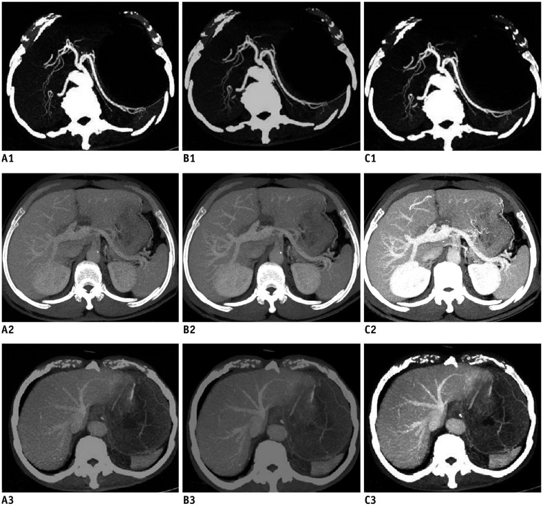
Fig. 1 Comparison of images.In phase of hepatic artery in QC (A1), in 50 keV (B1), and in 70 keV (C1). In phase of portal vein for QC (A2), in 50 keV (B2), and in 70 keV (C2). In phase of hepatic vein in QC (A3), in 50 keV (B3), and in 70 keV (C3). QC = quality check
Reference
-
1. Kishi Y, Imamura H, Sugawara Y, Sano K, Kaneko J, Kokudo N, et al. Evaluation of donor vasculobiliary anatomic variations in liver graft procurements. Surgery. 2010; 147:30–39. PMID: 19879610.
Article2. Radtke A, Sotiropoulos GC, Sgourakis G, Molmenti EP, Schroeder T, Saner FH, et al. Hepatic venous drainage: how much can we learn from imaging studies? Anatomic-functional classification derived from three-dimensional computed tomography reconstructions. Transplantation. 2010; 89:1518–1525. PMID: 20410853.
Article3. Radtke A, Sotiropoulos GC, Sgourakis G, Molmenti EP, Schroeder T, Saner FH, et al. “Anatomical” versus “territorial” belonging of the middle hepatic vein: virtual imaging and clinical repercussions. J Surg Res. 2011; 166:146–155. PMID: 19932902.
Article4. Takeishi K, Shirabe K, Yoshida Y, Tsutsui Y, Kurihara T, Kimura K, et al. Correlation between portal vein anatomy and bile duct variation in 407 living liver donors. Am J Transplant. 2015; 15:155–160. PMID: 25521764.
Article5. Goo HW, Goo JM. Dual-energy CT: new horizon in medical imaging. Korean J Radiol. 2017; 18:555–569. PMID: 28670151.
Article6. Song I, Yi JG, Park JH, Kim SM, Lee KS, Chung MJ. Virtual non-contrast CT using dual-energy spectral CT: feasibility of coronary artery calcium scoring. Korean J Radiol. 2016; 17:321–329. PMID: 27134521.
Article7. Xing Y, Zhao Y, Guo N, Pan CX, Azati G, Wang YW, et al. Effect of a novel intracycle motion correction algorithm on dual-energy spectral coronary CT angiography: a study with pulsating coronary artery phantom at high heart rates. Korean J Radiol. 2017; 18:881–887. PMID: 29089820.
Article8. Lv P, Lin XZ, Li J, Li W, Chen K. Differentiation of small hepatic hemangioma from small hepatocellular carcinoma: recently introduced spectral CT method. Radiology. 2011; 259:720–729. PMID: 21357524.
Article9. He J, Ma X, Wang Q, Fan J, Sun Z. Spectral CT demonstration of the superior mesenteric artery: comparison of monochromatic and polychromatic imaging. Acad Radiol. 2014; 21:364–368. PMID: 24360636.10. Xin L, Yang X, Huang N, Du X, Zhang J, Wang Y, et al. The initial experience of the upper abdominal CT angiography using low-concentration contrast medium on dual energy spectral CT. Abdom Imaging. 2015; 40:2894–2899. PMID: 26036793.
Article11. Zhao LQ, He W, Li JY, Chen JH, Wang KY, Tan L. Improving image quality in portal venography with spectral CT imaging. Eur J Radiol. 2012; 81:1677–1681. PMID: 21444170.
Article12. Fang CH, You JH, Lau WY, Lai EC, Fan YF, Zhong SZ, et al. Anatomical variations of hepatic veins: three-dimensional computed tomography scans of 200 subjects. World J Surg. 2012; 36:120–124. PMID: 21976007.
Article13. Wu TC, Lee RC, Chau GY, Chiang JH, Chang CY. Reappraisal of right portal segmental ramification based on 3-dimensional volume rendering of computed tomography during arterial portography. J Comput Assist Tomogr. 2007; 31:475–480. PMID: 17538299.
Article14. Zhao LQ, He W, Chen G. Characteristics of paraesophageal varices: a study with 64-row multidetector computed tomography portal venography. World J Gastroenterol. 2008; 14:5331–5335. PMID: 18785288.15. Coursey CA, Nelson RC, Boll DT, Paulson EK, Ho LM, Neville AM, et al. Dual-energy multidetector CT: how does it work, what can it tell us, and when can we use it in abdominopelvic imaging? Radiographics. 2010; 30:1037–1055. PMID: 20631367.
Article16. Cheng J, Yin Y, Wu H, Zhang Q, Hua J, Hua X, et al. Optimal monochromatic energy levels in spectral CT pulmonary angiography for the evaluation of pulmonary embolism. PLoS One. 2013; 8:e63140. PMID: 23667583.
Article17. Schueller-Weidekamm C, Schaefer-Prokop CM, Weber M, Herold CJ, Prokop M. CT angiography of pulmonary arteries to detect pulmonary embolism: improvement of vascular enhancement with low kilovoltage settings. Radiology. 2006; 241:899–907. PMID: 17114631.
Article18. Takahashi H, Okada M, Hyodo T, Hidaka S, Kagawa Y, Matsuki M, et al. Can low-dose CT with iterative reconstruction reduce both the radiation dose and the amount of iodine contrast medium in a dynamic CT study of the liver? Eur J Radiol. 2014; 83:684–691. PMID: 24418284.
Article19. Wang X, Zhong Y, Hu L, Xue L, Shi M, Qiu H, et al. A prospective evaluation of the contrast, radiation dose and image quality of contrast-enhanced CT scans of paediatric abdomens using a low-concentration iodinated contrast agent and low tube voltage combined with 70% ASIR algorithm. Int J Clin Pract. 2016; 70(Suppl 9B):B16–B21. PMID: 27577509.
Article20. Huda W, Scalzetti EM, Levin G. Technique factors and image quality as functions of patient weight at abdominal CT. Radiology. 2000; 217:430–435. PMID: 11058640.
Article21. Funama Y, Awai K, Nakayama Y, Kakei K, Nagasue N, Shimamura M, et al. Radiation dose reduction without degradation of low-contrast detectability at abdominal multisection CT with a low-tube voltage technique: phantom study. Radiology. 2005; 237:905–910. PMID: 16237139.
Article22. Nakayama Y, Awai K, Funama Y, Hatemura M, Imuta M, Nakaura T, et al. Abdominal CT with low tube voltage: preliminary observations about radiation dose, contrast enhancement, image quality, and noise. Radiology. 2005; 237:945–951. PMID: 16237140.
Article23. Wintersperger B, Jakobs T, Herzog P, Schaller S, Nikolaou K, Suess C, et al. Aorto-iliac multidetector-row CT angiography with low kV settings: improved vessel enhancement and simultaneous reduction of radiation dose. Eur Radiol. 2005; 15:334–341. PMID: 15611872.
Article24. Sagara Y, Hara AK, Pavlicek W, Silva AC, Paden RG, Wu Q. Abdominal CT: comparison of low-dose CT with adaptive statistical iterative reconstruction and routine-dose CT with filtered back projection in 53 patients. AJR Am J Roentgenol. 2010; 195:713–719. PMID: 20729451.
Article25. Ma CL, Chen XX, Lei YX, Zhang XR, Jia YJ, Tian X, et al. Clinical value of dual-energy spectral imaging with adaptive statistical iterative reconstruction for reducing contrast medium dose in CT portal venography: in comparison with standard 120-kVp imaging protocol. Br J Radiol. 2016; 89:20151022. PMID: 27031376.
Article26. Zhao Y, Wu Y, Zuo Z, Suo H, Zhao S, Han J, et al. Application of low concentration contrast medium in spectral CT imaging for CT portal venography. J Xray Sci Technol. 2017; 25:135–143. PMID: 27768006.
Article27. Zhu Z, Zhao XM, Zhao YF, Wang XY, Zhou CW. Feasibility study of using gemstone spectral imaging (GSI) and adaptive statistical iterative reconstruction (ASIR) for reducing radiation and iodine contrast dose in abdominal CT patients with high BMI values. PLoS One. 2015; 10:e0129201. PMID: 26079259.
Article
- Full Text Links
-
- Actions
-
Cited
- CITED
-
- Close
- Share
-
- Similar articles
-
- Dual-Energy Computed Tomography Arthrography of the Shoulder Joint Using Virtual Monochromatic Spectral Imaging: Optimal Dose of Contrast Agent and Monochromatic Energy Level
- Effect of a Novel Intracycle Motion Correction Algorithm on Dual-Energy Spectral Coronary CT Angiography: A Study with Pulsating Coronary Artery Phantom at High Heart Rates
- Preliminary Application of High-Definition CT Gemstone Spectral Imaging in Hand and Foot Tendons
- Application of Dual-Energy Spectral Computed Tomography to Thoracic Oncology Imaging
- Future of quality management of medical imaging
