The Safety of Protected Carotid Artery Stenting in Patients with Unstable Plaque on Carotid High-Resolution MR Imaging
- Affiliations
-
- 1Department of Radiology, Chonbuk National University Medical School and Hospital, Jeonju, Korea. kwak8140@jbnu.ac.kr
- 2Research Institute of Clinical Medicine of Chonbuk National University-Biomedical Research Institute of Chonbuk National University Hospital, Jeonju, Korea.
- KMID: 2412356
- DOI: http://doi.org/10.3348/jksr.2018.78.6.380
Abstract
- PURPOSE
The aim of this study was to determine the safety of the carotid artery stenting (CAS) procedure, using an embolic protection device which is based on the presence of unstable plaques on carotid MR imaging in patients who presented with severe carotid artery stenosis.
MATERIALS AND METHODS
This prospective study assessed 102 consecutive patients who had been noted with severe carotid stenosis. These patients underwent a preprocedural carotid MR imaging, and a periprocedural diffusion-weighted imaging (DWI) after the CAS. The unstable plaque on the carotid MR imaging was defined as characterized as intraplaque hemorrhage (IPH), thin/ruptured fibrous caps, or ulcers. We analyzed the incidence of the noted periprocedural ipsilateral ischemic events on the DWI, and noted the primary outcomes within 30 days of the CAS.
RESULTS
In the study, it is noted that 50 patients (49.0%) had IPH, 84 patients (82.4%) had thin/ruptured fibrous caps, and 43 patients (42.2%) had ulcers as seen on the carotid plaque MR imaging. The IPH was more common in the symptomatic group than in the asymptomatic group (58.7% vs. 41.1%, p = 0.12). Overall, the DWI was positive after CAS in 25.5% of cases. Additionally, the combined rate of 30-day stroke, myocardial infarction, or death was recorded at 3.9%. The new periprocedural ischemic lesions on the DWI were characteristically more frequently observed in the symptomatic group (17/46, 37.0%) than in the asymptomatic group (9/56, 16.1%) (p = 0.03). There was no significant difference in the primary outcome of the CAS, based on the type of unstable plaque of IPH, thin/ruptured fibrous caps, or ulcers.
CONCLUSION
The protected CAS appears to be safe, regardless of the noted unstable plaque findings as seen on the carotid MR imaging. In this case, because of the higher risk of periprocedural ipsilateral ischemic events, it is therefore recommended that the symptomatic patients should receive more careful treatment during the CAS placement going forward.
MeSH Terms
Figure
-
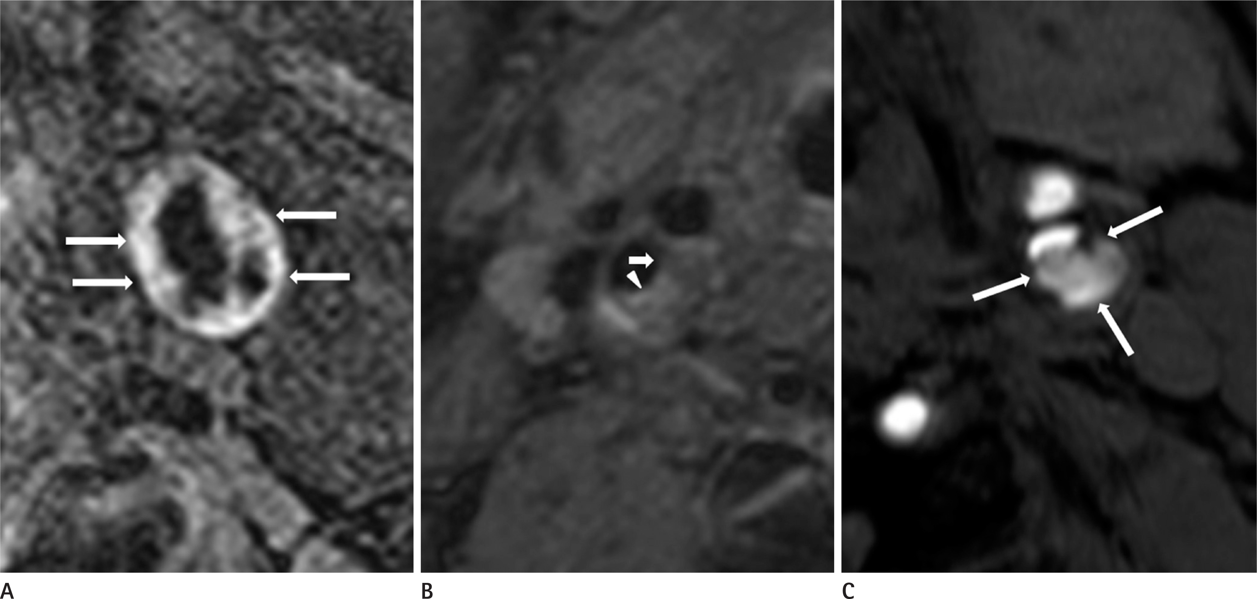
Fig. 1. Carotid MR imaging of unstable plaque. A. Intraplaque hemorrhage: presence of hyperintense signal within carotid plaque on carotid magnetization-prepared rapid acquisition with gradient-echo sequence (arrows). B. Thin/ruptured fibrous cap: no contrast enhancement (arrow) compared to the surrounding, more strongly enhanced fibrous cap on postcontrast T1-weighted imaging (arrowhead). C. Ulcer: depression below the plaque surface on carotid MR imaging (arrows: large penetrating ulcer).
Cited by 1 articles
-
Findings of Angiography and Carotid Vessel Wall Imaging Associated with Post-Procedural Clinical Events after Carotid Artery Stenting
Sujin Jeon, Heejae Park, Hyo Sung Kwak, Seung Bae Hwang
Neurointervention. 2024;19(1):14-23. doi: 10.5469/neuroint.2023.00486.
Reference
-
References
1. Virmani R, Burke AP, Kolodgie FD, Farb A. Vulnerable plaque: the pathology of unstable coronary lesions. J Interv Cardiol. 2002; 15:439–446.
Article2. Fuster V, Stein B, Ambrose JA, Badimon L, Badimon JJ, Che-sebro JH. Atherosclerotic plaque rupture and thrombosis. evolving concepts. Circulation. 1990; 82(3 Suppl):): II. 47–59.3. Cai J, Hatsukami TS, Ferguson MS, Kerwin WS, Saam T, Chu B, et al. In vivo quantitative measurement of intact fibrous cap and lipid-rich necrotic core size in atherosclerotic carotid plaque: comparison of high-resolution, contrast-enhanced magnetic resonance imaging and histology. Circulation. 2005; 112:3437–3444.4. Takaya N, Cai J, Ferguson MS, Yarnykh VL, Chu B, Saam T, et al. Intra- and interreader reproducibility of magnetic resonance imaging for quantifying the lipid-rich necrotic core is improved with gadolinium contrast enhancement. J Magn Reson Imaging. 2006; 24:203–210.
Article5. Cai JM, Hatsukami TS, Ferguson MS, Small R, Polissar NL, Yuan C. Classification of human carotid atherosclerotic lesions with in vivo multicontrast magnetic resonance imaging. Circulation. 2002; 106:1368–1373.
Article6. Clarke SE, Hammond RR, Mitchell JR, Rutt BK. Quantitative assessment of carotid plaque composition using multicontrast MRI and registered histology. Magn Reson Med. 2003; 50:1199–1208.
Article7. Saam T, Cai JM, Cai YQ, An NY, Kampschulte A, Xu D, et al. Carotid plaque composition differs between ethno-racial groups: an MRI pilot study comparing mainland Chinese and American Caucasian patients. Arterioscler Thromb Vasc Biol. 2005; 25:611–616.8. Cappendijk VC, Cleutjens KB, Kessels AG, Heeneman S, Sch-urink GW, Welten RJ, et al. Assessment of human atherosclerotic carotid plaque components with multisequence MR imaging: initial experience. Radiology. 2005; 234:487–492.
Article9. Takaya N, Yuan C, Chu B, Saam T, Underhill H, Cai J, et al. Association between carotid plaque characteristics and subsequent ischemic cerebrovascular events: a prospective assessment with MRI–initial results. Stroke. 2006; 37:818–823.
Article10. Singh N, Moody AR, Gladstone DJ, Leung G, Ravikumar R, Zhan J, et al. Moderate carotid artery stenosis: MR imaging-depicted intraplaque hemorrhage predicts risk of cerebrovascular ischemic events in asymptomatic men. Radiology. 2009; 252:502–508.
Article11. Altaf N, Daniels L, Morgan PS, Auer D, MacSweeney ST, Moody AR, et al. Detection of intraplaque hemorrhage by magnetic resonance imaging in symptomatic patients with mild to moderate carotid stenosis predicts recurrent neurological events. J Vasc Surg. 2008; 47:337–342.
Article12. Rothwell PM, Gibson R, Warlow CP. Interrelation between plaque surface morphology and degree of stenosis on carotid angiograms and the risk of ischemic stroke in patients with symptomatic carotid stenosis. on behalf of the European Carotid Surgery Trialists'Collaborative Group. Stroke. 2000; 31:615–621.13. Eliasziw M, Streifler JY, Fox AJ, Hachinski VC, Ferguson GG, Barnett HJ. Significance of plaque ulceration in symptomatic patients with high-grade carotid stenosis. North American Symptomatic Carotid Endarterectomy Trial. Stroke. 1994; 25:304–308.
Article14. Bonati LH, Dobson J, Featherstone RL, Ederle J, van der Worp HB, de Borst GJ, et al. Long-term outcomes after stenting versus endarterectomy for treatment of symptomatic carotid stenosis: the International Carotid Stenting Study (ICSS) randomised trial. Lancet. 2015; 385:529–538.
Article15. Brott TG, Hobson RW, Howard G, Roubin GS, Clark WM, Brooks W, et al. Stenting versus endarterectomy for treatment of carotid-artery stenosis. N Engl J Med. 2010; 363:11–23.
Article16. Grimm JC, Arhuidese I, Beaulieu RJ, Qazi U, Perler BA, Freischlag JA, et al. Surgeon's 30-day outcomes supporting the carotid revascularization endarterectomy versus stenting trial. JAMA Surg. 2014; 149:1314–1318.
Article17. Mantese VA, Timaran CH, Chiu D, Begg RJ, Brott TG. CREST Investigators. The Carotid Revascularization Endarterectomy versus Stenting Trial (CREST): stenting versus carotid endarterectomy for carotid disease. Stroke. 2010; 41(10 Suppl):): S31-S34.
Article18. Yoshimura S, Yamada K, Kawasaki M, Asano T, Kanematsu M, Takamatsu M, et al. High-intensity signal on time-of-flight magnetic resonance angiography indicates carotid plaques at high risk for cerebral embolism during stenting. Stroke. 2011; 42:3132–3137.
Article19. Yoon W, Kim SK, Park MS, Chae HJ, Kang HK. Safety of protected carotid artery stenting in patients with severe carotid artery stenosis and carotid intraplaque hemorrhage. AJNR Am J Neuroradiol. 2012; 33:1027–1031.
Article20. Chung GH, Jeong JY, Kwak HS, Hwang SB. Associations between cerebral embolism and carotid intraplaque hemorrhage during protected carotid artery stenting. AJNR Am J Neuroradiol. 2016; 37:686–691.
Article21. Yoshimura S, Yamada K, Kawasaki M, Asano T, Kanematsu M, Miyai M, et al. Selection of carotid artery stenting or endarterectomy based on magnetic resonance plaque imaging reduced periprocedural adverse events. J Stroke Cerebrovasc Dis. 2013; 22:1082–1087.22. Sun J, Balu N, Hippe DS, Xue Y, Dong L, Zhao X, et al. Subclinical carotid atherosclerosis: short-term natural history of lipid-rich necrotic core–a multicenter study with MR imaging. Radiology. 2013; 268:61–68.
Article23. Sun J, Underhill HR, Hippe DS, Xue Y, Yuan C, Hatsukami TS. Sustained acceleration in carotid atherosclerotic plaque progression with intraplaque hemorrhage: a longterm time course study. JACC Cardiovasc Imaging. 2012; 5:798–804.24. Lee KJ, Kwak HS, Chung GH, Hwang SB, Song JS. Assessment of carotid diffusion-weighted imaging for detection of lipid-rich necrotic core in symptomatic carotid atheroma. J Korean Soc Radiol. 2016; 74:160–168.
Article25. Zhao XQ, Dong L, Hatsukami T, Phan BA, Chu B, Moore A, et al. MR imaging of carotid plaque composition during lipid-lowering therapy a prospective assessment of effect and time course. JACC Cardiovasc Imaging. 2011; 4:977–986.26. Saam T, Hetterich H, Hoffmann V, Yuan C, Dichgans M, Pop-pert H, et al. Meta-analysis and systematic review of the predictive value of carotid plaque hemorrhage on cerebrovascular events by magnetic resonance imaging. J Am Coll Cardiol. 2013; 62:1081–1091.
Article27. Chiam PTL, Roubin GS, Iyer SS, Green RM, Soffer DE, Brennan C, et al. Carotid artery stenting in elderly patients: importance of case selection. Catheter Cardiovasc Interv. 2008; 72:318–324.
Article28. Bijuklic K, Wandler A, Varnakov Y, Tuebler T, Schofer J. Risk factors for cerebral embolization after carotid artery stenting with embolic protection: a diffusion-weighted magnetic resonance imaging study in 837 consecutive patients. Circ Cardiovasc Interv. 2013; 6:311–316.29. Schnaudigel S, Gröschel K, Pilgram SM, Kastrup A. New brain lesions after carotid stenting versus carotid endarterectomy: a systematic review of the literature. Stroke. 2008; 39:1911–1919.30. Yang M, Yu Y, Walsh WR, Yang JL, Baker L, Lennox AF, et al. A microscopic and biomarker evaluation of embolic filter debris collected during carotid artery stenting. J Endovasc Ther. 2016; 23:275–284.
Article
- Full Text Links
-
- Actions
-
Cited
- CITED
-
- Close
- Share
-
- Similar articles
-
- High-Resolusion Magnetic Resonance Imaging of Carotid Atherosclerotic Plaque
- Usefulness of carotid ultrasonography and treatment of carotid disease
- A Case of Transseptal Approach to Carotid Artery Stenting in Right Internal Carotid Stenosis
- Massive Cerebral Microemboli after Protected Carotid Artery Angioplasty and Stenting Using a Distal Filter Embolic Protection Device for a Vulnerable Plaque with a Lipid Rich Necrotic Core and Intraplaque Hemorrhage: A Case Report
- Relationship between Carotid Artery Plaque Measured by Ultrasound and Cerebral infarction in Patients with Non-insulin Dependent Diabetes
