A Prospective Blinded Study of Endoscopic Ultrasound Elastography in Liver Disease: Towards a Virtual Biopsy
- Affiliations
-
- 1Division of Gastroenterology, Hepatology and Endoscopy, Brigham and Women's Hospital, Boston, MA, USA. mryou@bwh.harvard.edu
- 2Department of Medicine, Harvard Medical School, Boston, MA, USA.
- KMID: 2410986
- DOI: http://doi.org/10.5946/ce.2017.095
Abstract
- BACKGROUND/AIMS
Liver biopsy has traditionally been used for determining the degree of fibrosis, however there are several limitations. Endoscopic ultrasound (EUS) real-time elastography (RTE) is a novel technology that uses image enhancement to display differences in tissue compressibility. We sought to assess whether liver fibrosis index (LFI) can distinguish normal, fatty, and cirrhotic liver tissue.
METHODS
A total of 50 patients undergoing EUS were prospectively enrolled. RTE of the liver was performed to synthesize the LFI in each patient. Univariate and multivariable analyses were performed. Chi-square and t-tests were performed for categorical and continuous variables, respectively. A p-value of <0.05 was considered significant.
RESULTS
Abdominal imaging prior to endoscopic evaluation suggested normal tissue, fatty liver, and cirrhosis in 26, 16, and 8 patients, respectively. Patients with cirrhosis had significantly increased mean LFI compared to the fatty liver (3.2 vs. 1.7, p<0.001) and normal (3.2 vs. 0.8, p<0.001) groups. The fatty liver group showed significantly increased LFI compared to the normal group (3.8 vs. 1.4, p<0.001). Multivariable regression analysis suggested that LFI was an independent predictor of group features (p<0.001).
CONCLUSIONS
LFI computed from RTE images significantly correlates with abdominal imaging and can distinguish normal, fatty, and cirrhotic-appearing livers; therefore, LFI may play an important role in patients with chronic liver disease.
Keyword
MeSH Terms
Figure
-
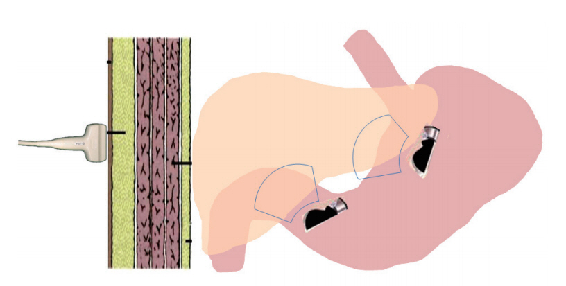
Fig. 1. Schematic demonstration of endoscopic ultrasound-based real time elastography, which can evaluate both the right and left liver through the gastrointestinal wall compared to trans-abdominal elastography, which requires signal transmission through the thick abdominal wall.
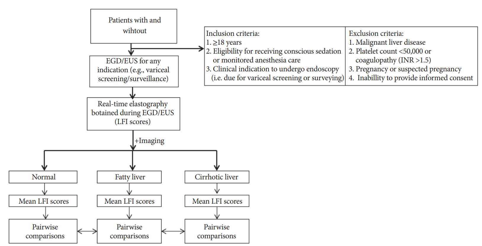
Fig. 2. Flowchart showing the basic study design. EGD, esophagogastroduodenoscopy; EUS, endoscopic ultrasound; LFI, liver fibrosis index; INR, international normalized ratio.
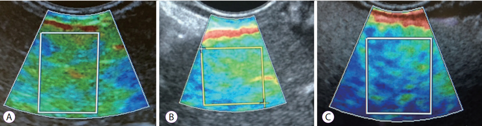
Fig. 3. Representative endoscopic ultrasound real-time elastography images in patients with normal (A), fatty (B), and cirrhotic (C) liver tissue on cross-sectional imaging.
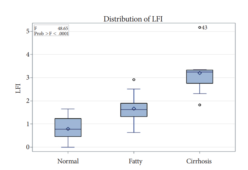
Fig. 4. Comparison of liver fibrosis index (LFI) scores between the normal, fatty, and cirrhotic liver imaging (ANOVA).
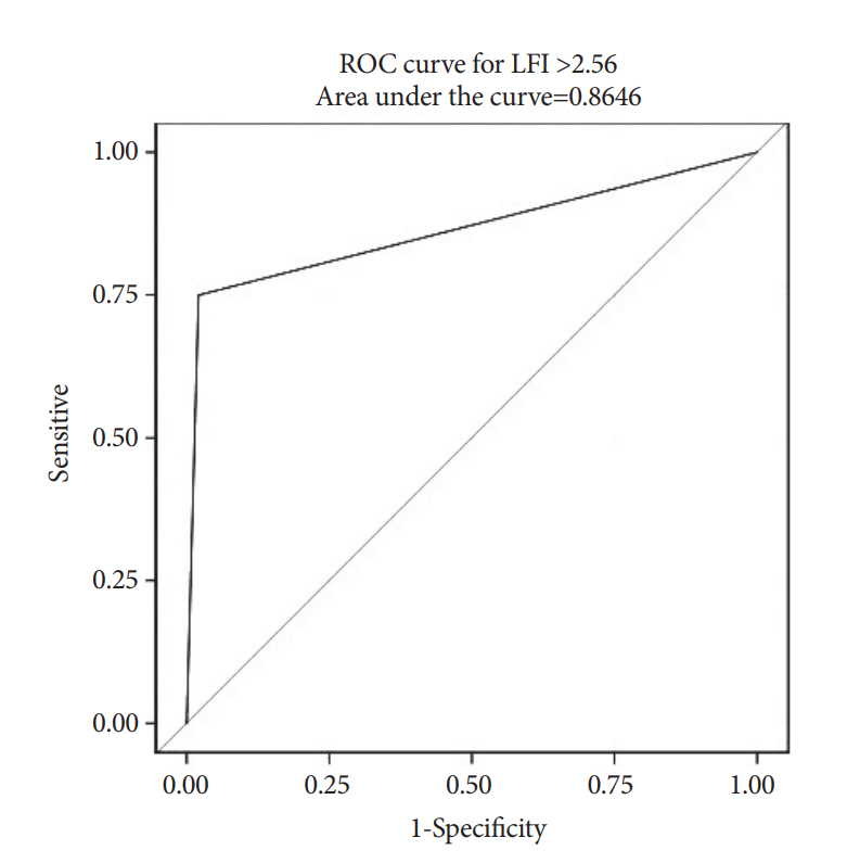
Fig. 5. Area under the receiver operator characteristic (ROC) curve for the prediction of cirrhosis on imaging by liver fibrosis index (LFI).
Cited by 1 articles
-
Endoscopic Ultrasound Real-Time Elastography in Liver Disease
Jeong Eun Song, Dong Wook Lee, Eun Young Kim
Clin Endosc. 2018;51(2):118-119. doi: 10.5946/ce.2018.049.
Reference
-
1. Bedossa P, Poynard T. An algorithm for the grading of activity in chronic hepatitis C. The METAVIR cooperative study group. Hepatology. 1996; 24:289–293.2. Pang JX, Zimmer S, Niu S, et al. Liver stiffness by transient elastography predicts liver-related complications and mortality in patients with chronic liver disease. PLoS One. 2014; 9:e95776.
Article3. Bedossa P, Dargère D, Paradis V. Sampling variability of liver fibrosis in chronic hepatitis C. Hepatology. 2003; 38:1449–1457.
Article4. Castera L, Forns X, Alberti A. Non-invasive evaluation of liver fibrosis using transient elastography. J Hepatol. 2008; 48:835–847.
Article5. Berzigotti A, Ashkenazi E, Reverter E, Abraldes JG, Bosch J. Non-invasive diagnostic and prognostic evaluation of liver cirrhosis and portal hypertension. Dis Markers. 2011; 31:129–138.
Article6. Augustin S, Millán L, González A, et al. Detection of early portal hypertension with routine data and liver stiffness in patients with asymptomatic liver disease: a prospective study. J Hepatol. 2014; 60:561–569.
Article7. Fraquelli M, Rigamonti C, Casazza G, et al. Reproducibility of transient elastography in the evaluation of liver fibrosis in patients with chronic liver disease. Gut. 2007; 56:968–973.
Article8. Castéra L, Foucher J, Bernard PH, et al. Pitfalls of liver stiffness measurement: a 5-year prospective study of 13,369 examinations. Hepatology. 2010; 51:828–835.
Article9. Wong GL, Wong VW, Chim AM, et al. Factors associated with unreliable liver stiffness measurement and its failure with transient elastography in the Chinese population. J Gastroenterol Hepatol. 2011; 26:300–305.
Article10. Chang PE, Goh GB, Ngu JH, Tan HK, Tan CK. Clinical applications, limitations and future role of transient elastography in the management of liver disease. World J Gastrointest Pharmacol Ther. 2016; 7:91–106.
Article11. Kettaneh A, Marcellin P, Douvin C, et al. Features associated with success rate and performance of fibroscan measurements for the diagnosis of cirrhosis in HCV patients: a prospective study of 935 patients. J Hepatol. 2007; 46:628–634.
Article12. Lucidarme D, Foucher J, Le Bail B, et al. Factors of accuracy of transient elastography (fibroscan) for the diagnosis of liver fibrosis in chronic hepatitis C. Hepatology. 2009; 49:1083–1089.
Article13. Tatsumi C, Kudo M, Ueshima K, et al. Non-invasive evaluation of hepatic fibrosis for type C chronic hepatitis. Intervirology. 2010; 53:76–81.
Article14. Robic MA, Procopet B, Métivier S, et al. Liver stiffness accurately predicts portal hypertension related complications in patients with chronic liver disease: a prospective study. J Hepatol. 2011; 55:1017–1024.
Article15. Tatsumi C, Kudo M, Ueshima K, et al. Noninvasive evaluation of hepatic fibrosis using serum fibrotic markers, transient elastography (fibroscan) and real-time tissue elastography. Intervirology. 2008; 51(Suppl 1):27–33.
Article
- Full Text Links
-
- Actions
-
Cited
- CITED
-
- Close
- Share
-
- Similar articles
-
- Ultrasound-based Liver Elastography: Recent Advances
- Elastography of the Pancreas, Current View
- Prospective Comparison of Liver Stiffness Measurements between Two Point Shear Wave Elastography Methods: Virtual Touch Quantification and Elastography Point Quantification
- Principles and clinical application of ultrasound elastography for diffuse liver disease
- Ultrasound Elastography in Differential Diagnosis of Benign and Malignant Thyroid Nodules
