Ann Surg Treat Res.
2018 May;94(5):274-278. 10.4174/astr.2018.94.5.274.
Primary malignant melanoma of the small intestine: a report of 2 cases and a review of the literature
- Affiliations
-
- 1Department of General Surgery, University of Ulsan College of Medicine, Gangneung Asan Hospital, Gangneung, Korea.
- 2Division of Colon and Rectal Surgery, Department of Surgery, Asan Medical Center, University of Ulsan College of Medicine, Seoul, Korea. jckim@amc.seoul.kr
- 3Department of Pathology, Asan Medical Center,University of Ulsan College of Medicine, Seoul, Korea.
- KMID: 2410272
- DOI: http://doi.org/10.4174/astr.2018.94.5.274
Abstract
- The majority of malignant melanomas in the small intestine are metastases from primary cutaneous lesions, it can also develop as a primary mucosal tumor in the gastrointestinal tract. In this report, we present rare cases of primary small bowel melanoma and review the current literature. A 78-year-old male presented with abdominal pain and CT enterography identified a ileal mass. A 79-year-old female presented with signs and symptoms of partial small bowel obstruction. Abdominopelvic CT and small bowel series revealed a obstructing mass in the distal jejunum. The masses were confirmed on laparotomy and histologically diagnosed as melanoma. Extensive postoperative clinical examination revealed no cutaneous lesions. A primary small bowel melanoma is an extremely rare neoplasm. A definite diagnosis can only be made after a thorough investigation has been made to exclude the coexistence of a primary lesion. Curative resection of the tumor remains the treatment of choice.
Keyword
MeSH Terms
Figure
-

Fig. 1 Surgical specimen (A, case 1; C, case 2) and macroscopic image of the tumor cut in half (B, case 1; D, case 2).
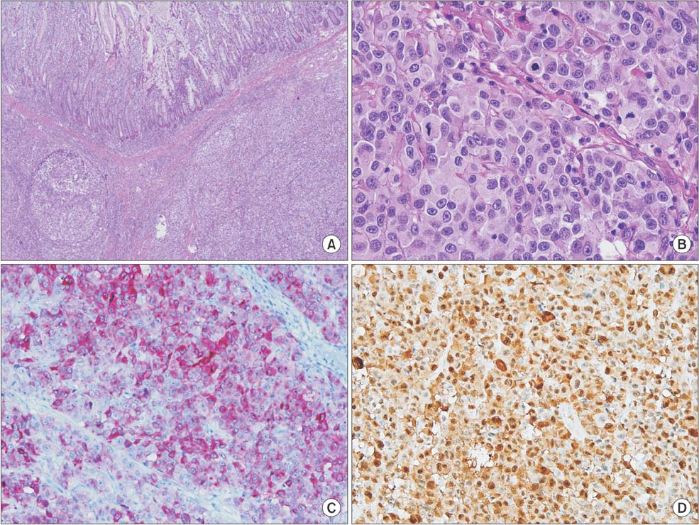
Fig. 2 Histologic feature of malignant melanoma. (A) The lesion shows diffuse atypical cells infiltration forming solid sheet (H&E, ×40). (B) The cells are medium-to large-sized with irregular nuclear contours, macronucleoli, and numerous mitotic activities (×400). Immunohistochemical staining showed that the malignant cells were strongly positive for Melan A (C) and S100 (D) (×200).
Reference
-
1. Blecker D, Abraham S, Furth EE, Kochman ML. Melanoma in the gastrointestinal tract. Am J Gastroenterol. 1999; 94:3427–3433.
Article2. Cheung MC, Perez EA, Molina MA, Jin X, Gutierrez JC, Franceschi D, et al. Defining the role of surgery for primary gastrointestinal tract melanoma. J Gastrointest Surg. 2008; 12:731–738.
Article3. Lens M, Bataille V, Krivokapic Z. Melanoma of the small intestine. Lancet Oncol. 2009; 10:516–521.
Article4. Hadjinic ol, Hadji t, Athanasopoulos PG, Shah R, Ala AA. Primary small bowel melanomas: fact or myth? Ann Transl Med. 2016; 4:113.5. Byrne EH, Fisher DE. Immune and molecular correlates in melanoma treated with immune checkpoint blockade. Cancer. 2017; 123(S11):2143–2153.
Article6. Spiridakis KG, Polichronaki EE, Sfakianakis EE, Flamourakis ME, Margetousakis TH, Xekalou AS, et al. Primary small bowel melanoma. A case report and a review of the literature. G Chir. 2015; 36:128–132.
Article7. Manouras A, Genetzakis M, Lagoudianakis E, Markogiannakis H, Papadima A, Kafiri G, et al. Malignant gastrointestinal melanomas of unknown origin: should it be considered primary? World J Gastroenterol. 2007; 13:4027–4029.
Article8. Timmers TK, Schadd EM, Monkelbaan JF, Meij V. Survival after resection of a primary malignant melanoma of the small intestine in a young patient: report of a case. Case Rep Gastroenterol. 2013; 7:251–260.
Article9. Khosrowshahi E, Horvath W. Primary malignant melanoma of the small intestine--a case report. Rontgenpraxis. 2002; 54:220–223.10. Iijima S, Oka K, Sasaki M, Tateishi Y, Saito H, Sandoh N, et al. Primary jejunal malignant melanoma first noticed because of the presence of parotid lymph node metastasis. J Am Acad Dermatol. 2003; 49:319–323.
Article11. Resta G, Anania G, Messina F, de Tullio D, Ferrocci G, Zanzi F, et al. Jejuno-jejunal invagination due to intestinal melanoma. World J Gastroenterol. 2007; 13:310–312.
Article
- Full Text Links
-
- Actions
-
Cited
- CITED
-
- Close
- Share
-
- Similar articles
-
- Primary Malignant Laryngeal Melanoma: Report of a Case with Review of Literature
- A Case of Primary Malignant Melanoma of the Vagina: Trial of a Wide Local Excision of Vagina and Rectum
- Primary Malignant Lymphoma of the Small Intestine Causing Adult Intussusception as an Initial Symptom
- A Case of Primary Small Bowel Melanoma Diagnosed by Single-Balloon Enteroscopy
- Esophagus, Stomach & Intestine; A Case of Primary Malignant Melanoma of the Esophagus
