J Korean Acad Prosthodont.
2018 Apr;56(2):173-178. 10.4047/jkap.2018.56.2.173.
Implant-supported prosthetic rehabilitation for the edentulous maxilla using the additive manufacturing technology: A case report
- Affiliations
-
- 1Department of Dentistry, Ajou University School of Medicine, Suwon, Republic of Korea. denthk@naver.com
- KMID: 2410122
- DOI: http://doi.org/10.4047/jkap.2018.56.2.173
Abstract
- The direct metal laser sintering (DMLS) technique would be promising for the full-arch implant-supported restorations due to reduced cost and manufacturing time without potential human errors and casting defects. The aims of this case report were to describe the successful outcome of an implant-supported fixed dental prosthesis in the edentulous maxilla by using the DMLS technology and computer-aided design and computer-aided manufacturing (CAD/CAM) monolithic zirconia crowns, and to describe its clinical implications. A healthy 51-year-old Korean woman visited Seoul National University Dental Hospital and she was in need of a rehabilitation of her entire maxilla due to severe tooth mobility. In this case, all maxillary teeth were extracted and an implant-supported fixed dental prosthesis was fabricated that involved a cobalt-chromium (Co-Cr) framework with the DMLS technique and CAD/CAM monolithic zirconia crowns. Six months after delivery, no distinct mechanical and biological complications were detected and the prosthesis exhibited satisfactory esthetics and function. In this case report, with the DMLS system, the three-dimensional printed prosthesis was created without additional manual tooling and thus, reliable accuracy and passive fit were obtained.
Keyword
MeSH Terms
Figure
-

Fig. 1 Initial clinical photo and radiograph. (A) Frontal view, (B) Panoramic radiograph.

Fig. 2 An intra-oral occlusal record was taken on the verification stent.

Fig. 3 The CAD model of a one-piece framework with screw access holes.
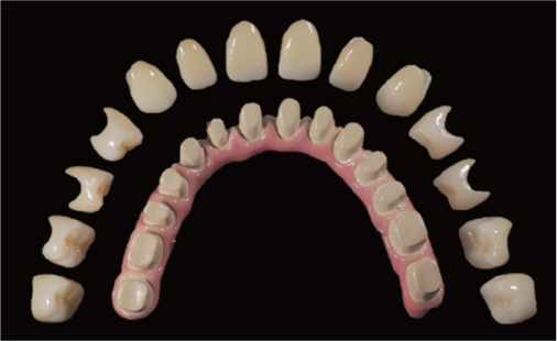
Fig. 4 Fourteen individual monolithic zirconia crowns were cemented on the Cobalt-chromium framework covered with porcelains.
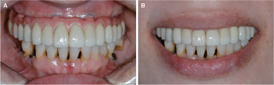
Fig. 5 Frontal views of the definitive prosthesis. (A) Maximum intercuspation position, (B) Posed smile.
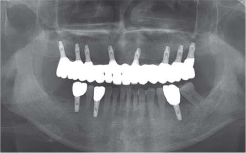
Fig. 6 Panoramic radiograph 6 months after delivery of the maxillary prosthesis.
Reference
-
1. van Noort R. The future of dental devices is digital. Dent Mater. 2012; 28:3–12.
Article2. Barazanchi A, Li KC, Al-Amleh B, Lyons K, Waddell JN. Additive technology: Update on current materials and applications in dentistry. J Prosthodont. 2017; 26:156–163.
Article3. Ploch CC, Mansi CSSA, Jayamohan J, Kuhl E. Using 3D printing to create personalized brain models for neurosurgical training and preoperative planning. World Neurosurg. 2016; 90:668–674.
Article4. Mangano C, De Rosa A, Desiderio V, d’Aquino R, Piattelli A, De Francesco F, Tirino V, Mangano F, Papaccio G. The osteoblastic differentiation of dental pulp stem cells and bone formation on different titanium surface textures. Biomaterials. 2010; 31:3543–3551.
Article5. Ge Z, Jin Z, Cao T. Manufacture of degradable polymeric scaffolds for bone regeneration. Biomed Mater. 2008; 3:022001.
Article6. Santos EC, Shiomi M, Osakada K, Laoui T. Rapid manufacturing of metal components by laser forming. Int J Mach Tools Manufact. 2006; 46:1459–1468.
Article7. Anusavice KJ, Shen C, Rawls HR. Phillip's science of dental materials. 12th ed. St. Louis: Elsevier;2012. p. 367–395.8. Tallgren A. The reduction in face height of edentulous and partially edentulous subjects during long-term denture wear. A longitudinal roentgenographic cephalometric study. Acta Odontol Scand. 1966; 24:195–239.
Article9. Jivraj S, Chee W, Corrado P. Treatment planning of the edentulous maxilla. Br Dent J. 2006; 201:261–279.
Article10. Sedda M, Vichi A, Carrabba M, Capperucci A, Louca C, Ferrari M. Influence of coloring procedure on flexural resistance of zirconia blocks. J Prosthet Dent. 2015; 114:98–102.
Article11. Kim HK, Kim SH. Optical properties of pre-colored dental monolithic zirconia ceramics. J Dent. 2016; 55:75–81.
Article12. Miller PD Jr. A classification of marginal tissue recession. Int J Periodontics Restorative Dent. 1985; 5:8–13.13. Schropp L, Wenzel A, Kostopoulos L, Karring T. Bone healing and soft tissue contour changes following single-tooth extraction: a clinical and radiographic 12-month prospective study. Int J Periodontics Restorative Dent. 2003; 23:313–323.14. Tjan AH, Miller GD, The JG. Some esthetic factors in a smile. J Prosthet Dent. 1984; 51:24–28.
Article15. Palmqvist S, Sondell K, Swartz B. Implant-supported maxillary overdentures: outcome in planned and emergency cases. Int J Oral Maxillofac Implants. 1994; 9:184–190.16. Ma S, Fenton A. Screw- versus cement-retained implant prostheses: a systematic review of prosthodontic maintenance and complications. Int J Prosthodont. 2015; 28:127–145.
Article17. Xu D, Xiang N, Wei B. The marginal fit of selective laser melting-fabricated metal crowns: an in vitro study. J Prosthet Dent. 2014; 112:1437–1440.
Article18. Örtorp A, Jönsson D, Mouhsen A, Vult von. The fit of cobalt-chromium three-unit fixed dental prostheses fabricated with four different techniques: a comparative in vitro study. Dent Mater. 2011; 27:356–363.
Article19. Tamac E, Toksavul S, Toman M. Clinical marginal and internal adaptation of CAD/CAM milling, laser sintering, and cast metal ceramic crowns. J Prosthet Dent. 2014; 112:909–913.
Article20. McLean JW, von Fraunhofer JA. The estimation of cement film thickness by an in vivo technique. Br Dent J. 1971; 131:107–111.
Article21. Kan JY, Rungcharassaeng K, Bohsali K, Goodacre CJ, Lang BR. Clinical methods for evaluating implant framework fit. J Prosthet Dent. 1999; 81:7–13.
Article
- Full Text Links
-
- Actions
-
Cited
- CITED
-
- Close
- Share
-
- Similar articles
-
- Rehabilitation of edentulous maxilla with implant-supported milled bar overdenture using CAD/CAM customized abutment: A case report
- A case of removable partial denture restoration using implant supported surveyed crown in a maxillary edentulous patient
- Full mouth rehabilitation of fully edentulous patient with implant-supported fixed prosthesis preceding bone graft: A case report
- Implant-supported overdenture manufactured using CAD/CAM techniques to achieve horizontal path insertion between the primary and secondary structure: A clinical case report
- Comparative evaluation of the subtractive and additive manufacturing on the color stability of fixed provisional prosthesis materials
