Factors Affecting Posterior Angulation in Retrograde Intramedullary Nailing for Distal Femoral Fractures
- Affiliations
-
- 1Department of Orthopedic Surgery, Cheju Halla General Hospital, Jeju, Korea. jihojeong76@gmail.com
- KMID: 2409760
- DOI: http://doi.org/10.12671/jkfs.2018.31.2.50
Abstract
- PURPOSE
To analyze the factors that cause a posterior angulatory deformity in the retrograde intramedullary nailing of distal femoral fractures.
MATERIALS AND METHODS
Fifty-five patients with distal femur fractures who were treated with retrograde intramedullary nailing were enrolled in this study. They were followed-up for at least one year postoperatively. The posterior angulatory deformity was evaluated according to the fracture location, pattern, and insertion point and the insertion point was compared with the ideal point derived from the radiographs of 100 normal adults. The correlation between the posterior angulation and the entry point of the nail was analyzed.
RESULTS
The posterior angulation was similar in terms of the fracture location; a meaningful difference was noted among the fracture patterns (p=0.047). The posterior angulation was significantly greater when the entry point was located more posteriorly, accepting a malreduced state (p=0.012).
CONCLUSION
Posterior angulation was smaller in the transverse fracture and the posterior location of the entry point from the apex of the Blumensaat's line increased the posterior angulation.
MeSH Terms
Figure
-
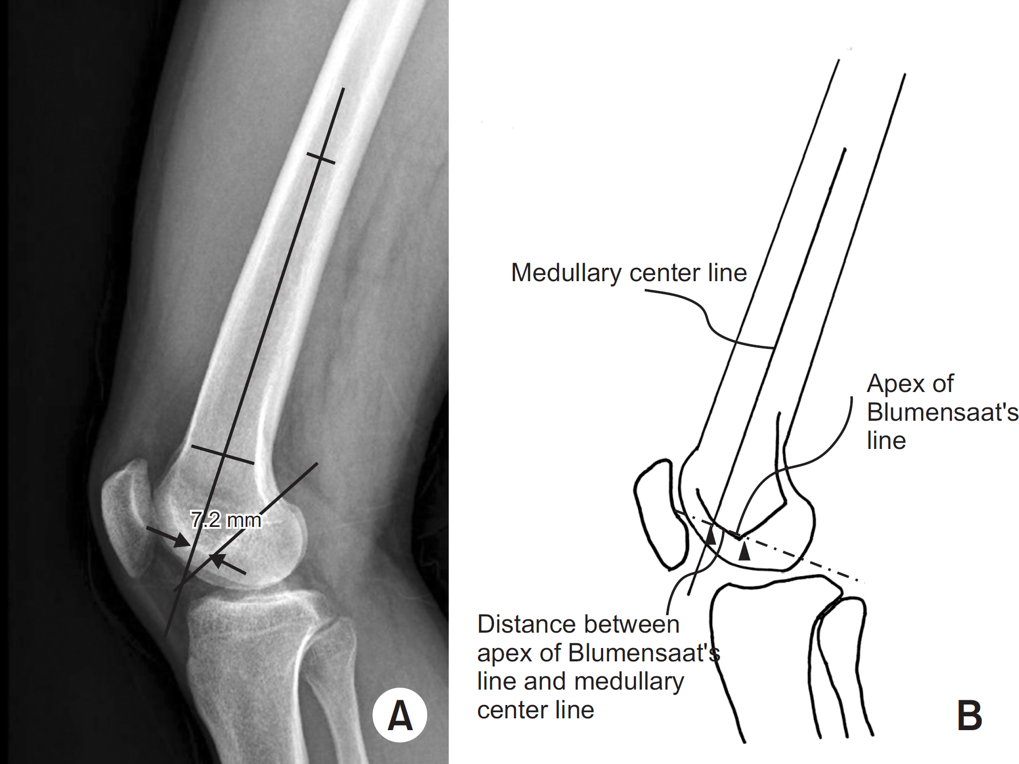
Fig. 1. Method of measuring the distance between the intramedullary center line and apex of the Blumensaat's line: (A) practical measurement, (B) schematic drawing.

Fig. 2. Entry point of the retrograde intramedullary nail, which could affect the posterior angulation of the fracture: (A) The 8.16 mm anterior location of entry point with no posterior angulation of fracture site was observed. (B) The 7.81 mm posterior location of entry point with posterior angulation.
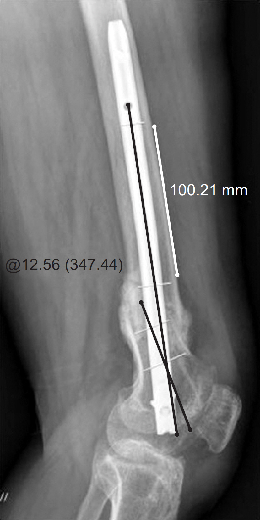
Fig. 3. Method for measuring the posterior angulation of the fracture site: That could be measured from the angle between an intramedullary center line of the distal fragment and that of the proximal fragment. This patient had malunion of 12.6o posterior angulation.

Fig. 4. Fracture level as a factor that could affect the posterior angulation of the fracture: (A) supracondylar level (AO 33 type), (B) distal one third level of femoral shaft (AO 32 type).
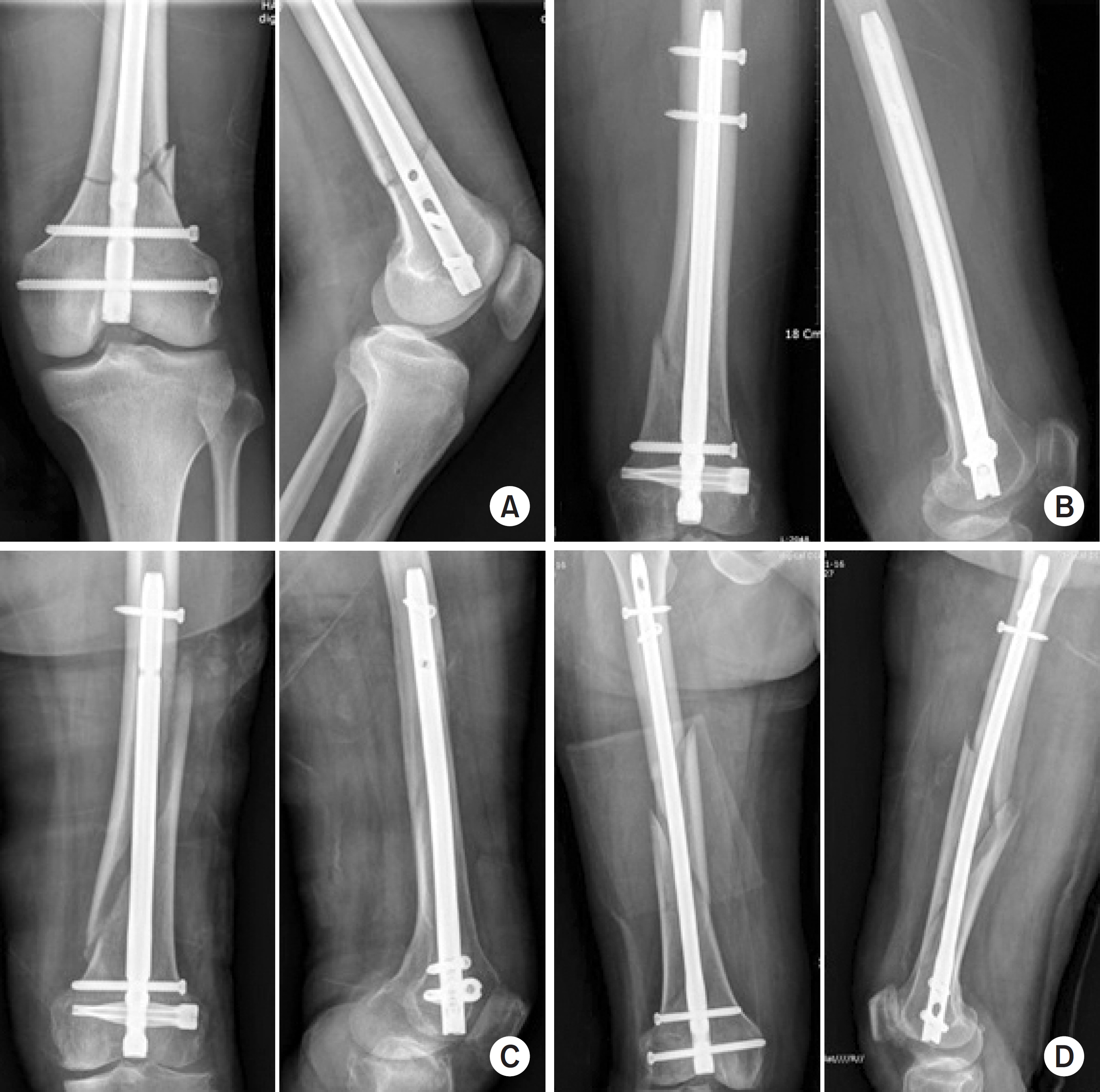
Fig. 5. Facture pattern as a factor that could affect the posterior angulation of a fracture: (A) transverse type, (B) oblique type, (C) spiral type, and (D) comminuted type.
Reference
-
References
1. Karpos PA, McFerran MA, Johnson KD. Intramedullary nailing of acute femoral shaft fractures using manual traction without a fracture table. J Orthop Trauma. 9:57–62. 1995.
Article2. Kempf I, Grosse A, Beck G. Closed locked intramedullary nailing. Its application to comminuted fractures of the femur. J Bone Joint Surg Am. 67:709–720. 1985.
Article3. Majkowski RS, Baker AS. Interlocking nails for femoral fractures: an initial experience. Injury. 22:93–96. 1991.
Article4. Wolinsky PR, McCarty E, Shyr Y, Johnson K. Reamed intramedullary nailing of the femur: 551 cases. J Trauma. 46:392–399. 1999.5. Moed BR, Watson JT. Retrograde intramedullary nailing, without reaming, of fractures of the femoral shaft in multiply injured patients. J Bone Joint Surg Am. 77:1520–1527. 1995.
Article6. Moed BR, Watson JT, Cramer KE, Karges DE, Teefey JS. Un-reamed retrograde intramedullary nailing of fractures of the femoral shaft. J Orthop Trauma. 12:334–342. 1998.
Article7. Patterson BM, Routt ML Jr, Benirschke SK, Hansen ST Jr. Retrograde nailing of femoral shaft fractures. J Trauma. 38:38–43. 1995.
Article8. Sanders R, Koval KJ, DiPasquale T, Helfet DL, Frankle M. Retrograde reamed femoral nailing. J Orthop Trauma. 7:293–302. 1993.
Article9. Swiontkowski MF, Hansen ST Jr, Kellam J. Ipsilateral fractures of the femoral neck and shaft. J Bone Joint Surg Am. 66:206–268. 1984.10. Ricci WM, Bellabarba C, Lewis R, et al. Angular malalignment after intramedullary nailing of femoral shaft fractures. J Orthop Trauma. 15:90–95. 2001.
Article11. Acharya KN, Rao MR. Retrograde nailing for distal third femoral shaft fractures: a prospective study. J Orthop Surg (Hong Kong). 14:253–258. 2006.
Article12. Ostrum RF. Treatment of floating knee injuries through a single percutaneous approach. Clin Orthop Relat Res. 375:43–50. 2000.
Article13. Whittle AP. Fractures of the lower extremity. Canale ST, Beaty JH, editors. Campbell's operative orthopaedics. 11th ed.Philadelphia, Mosby Elsevier: 3085–3236;2008.
Article14. O'Brien PJ, Meek RN, Blachut PA, Broekhuyse H. Fractures of the distal femur. Bucholz RW, Heckman JD, Court-Brown CM, editors. Rockwood and Green's fractures in adults. 6th ed.Philadelphia, Lippincott Williams & Wilkins: 1915–1967;2006.15. Gozna ER. Biomechanics of long bone injuries. Gonza ER, Harrington IJ, Evans DC, editors. Biomechanics of musculoskeletal injury. Baltimore: Williams & Wilkins;p. 1–29. 1982.16. Krupp RJ, Malkani AL, Goodin RA, Voor MJ. Optimal entry point for retrograde femoral nailing. J Orthop Trauma. 17:100–105. 2003.
Article17. Carmack DB, Moed BR, Kingston C, Zmurko M, Watson JT, Richardson M. Identification of the optimal intercondylar starting point for retrograde femoral nailing: an anatomic study. J Trauma. 55:692–695. 2003.
Article
- Full Text Links
-
- Actions
-
Cited
- CITED
-
- Close
- Share
-
- Similar articles
-
- Factors Affecting the Period of Bone Union When Treating Femoral Fractures with a Retrograde Intramedullary Nail
- Treatment of Distal Femoral Shaft and Supracondylar Fracture with aRetrograde Intramedullary Nailing
- Interlocking Intramedullary Nailing Versus conventional Kuntscher Intramedullary Nailing for Fracture of the Femoral Shaft
- Treatment of Distal Femoral Fractures with a Retrograde Supracondylar Intramedullary Nail assisted with Arthroscopy
- Closed Interlocking Nailing for Femoral Shaft Fracture -Comparison of results according to fracture comminution and site-
