Estragole Exhibits Anti-inflammatory Activity with the Regulation of NF-κB and Nrf-2 Signaling Pathways in LPS-induced RAW 264.7 cells
- Affiliations
-
- 1Department of Food and Life Science, Pukyong National University, Busan 608-737, Republic of Korea. choijs@pknu.ac.kr
- 2Department of Pharmaceutical Engineering, Sangji University, Wonju 220-702, Republic of Korea.
- 3Department of Food Science and Human Nutrition, Chonbuk National University, Jeonju 561-756, Republic of Korea. jungha@jbnu.ac.kr
- KMID: 2409604
- DOI: http://doi.org/10.20307/nps.2018.24.1.13
Abstract
- Estragole is a naturally occurring phenylpropanoid obtained from essential oils found in a broad diversity of plants. Although the phenylpropanoids show many biological activities, clear regulation of the inflammatory signaling pathways has not yet been determined. Here, we scrutinized the anti-inflammatory effect of estragole. The anti-inflammatory effect of estragole was determined through the inhibitory mechanisms of inducible nitric oxide synthase (iNOS), cyclooxygenase (COX-2), nuclear factor kappa B (NF-κB), and mitogen-activated protein kinases (MAPK) pathways and the activation of nuclear factor erythroid 2-related factor 2 (Nrf-2)/heme oxygenase (HO)-1 pathways in lipopolysaccharide (LPS)-stimulated RAW 264.7 cells. Estragole significantly inhibited NO production, iNOS and COX-2 expression as well as LPS-induced NF-κB and MAPK activation. Furthermore, estragole suppressed LPS-induced intracellular ROS production but up-regulated the stress response gene HO-1 via the activation of transcription factor Nrf-2. These findings demonstrate that estragole inhibits the LPS-induced expression of inflammatory mediators via the down-regulation of iNOS, COX-2, NF-κB, and MAPK pathways, as well as the up-regulation of the Nrf-2/HO-1 pathway, indicating that this phenylpropanoid has potential therapeutic and preventive applications in various inflammatory diseases.
Keyword
MeSH Terms
-
Down-Regulation
Mitogen-Activated Protein Kinases
NF-kappa B
Nitric Oxide Synthase Type II
Oils, Volatile
Prostaglandin-Endoperoxide Synthases
RAW 264.7 Cells*
Transcription Factors
Up-Regulation
Mitogen-Activated Protein Kinases
NF-kappa B
Nitric Oxide Synthase Type II
Oils, Volatile
Prostaglandin-Endoperoxide Synthases
Transcription Factors
Figure
-
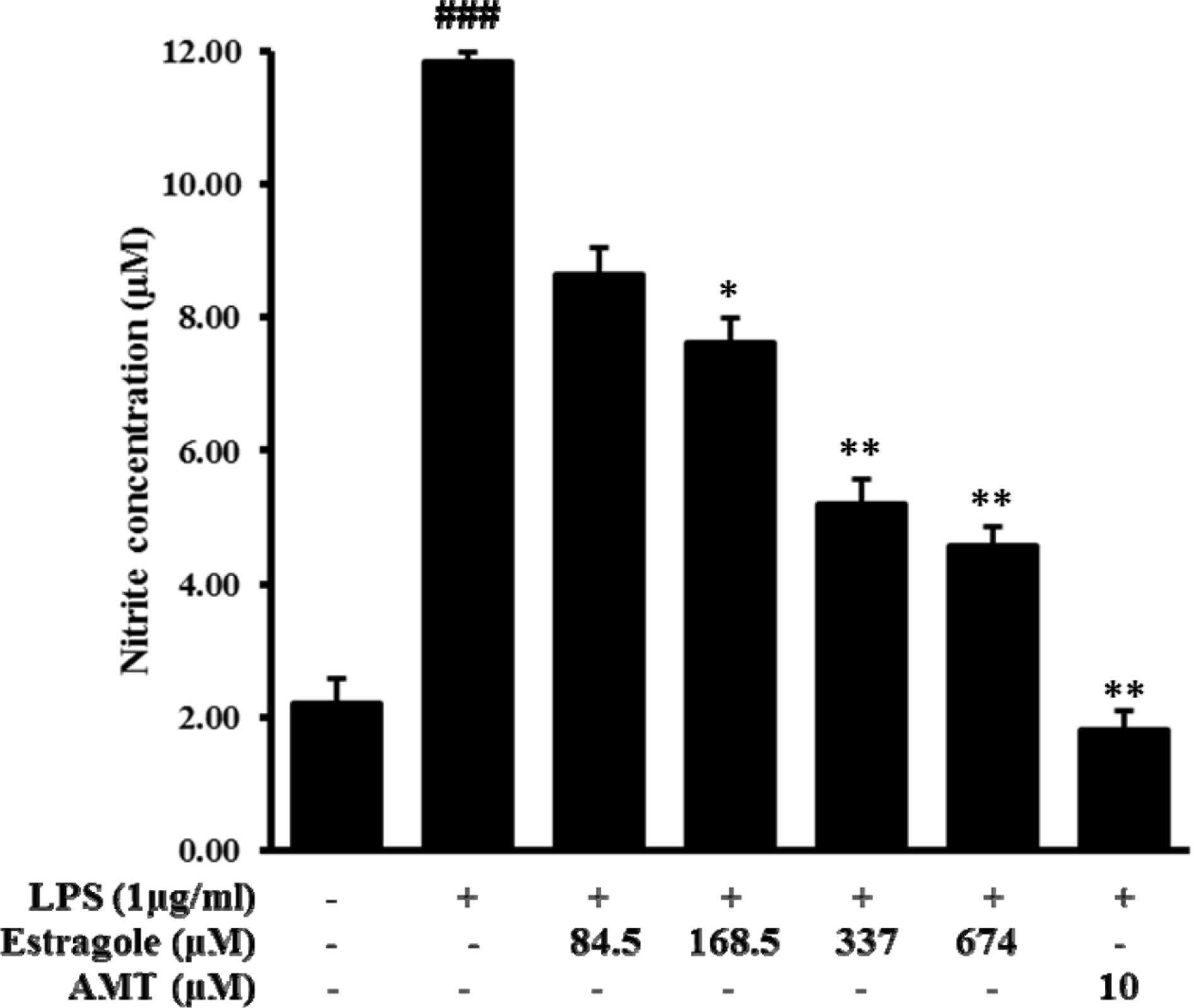
Fig. 1. Inhibitory effect of estragole on the production of nitric oxide (NO) in LPS-stimulated RAW264.7 cells. Cells were pretreated with different concentrations of estragole for 2 h and stimulated with LPS (1 μg/mL) for 24 h. NO production was measured by Griess reaction. Data are presented as the mean ± standard deviation of three independent experiments.###p < 0.001 indicates significant differences from the control group. ∗p <0.05, ∗∗p <0.01, and ∗∗∗p <0.001 indicate significant differences from the LPS-treated group.
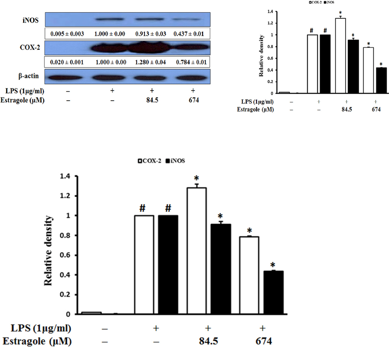
Fig. 2. Inhibitory effect of estragole on the expression of iNOS and COX-2 in LPS-stimulated RAW264.7 cells. Cells were pretreated with the indicated concentration of estragole for 2 h and stimulated with LPS (1 μg/mL) for 18 h. Data was detected by Western blot analysis with the designated antibodies. β-Actin was used as an internal control. The results presented are representative of three independent experiments. #p < 0.05 indicates significant differences from the control group. ∗p < 0.05 indicates significant differences from the LPS-treated group.
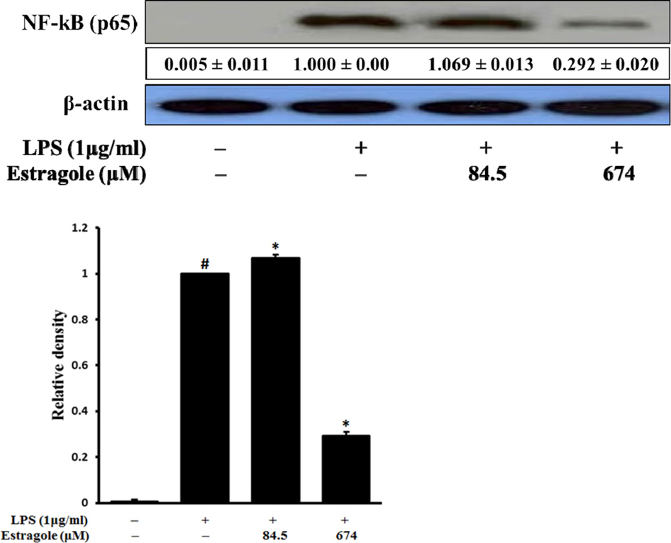
Fig. 3. Inhibitory effect of estragole on the expression of NF-κB (total protein) in LPS-stimulated RAW264.7 cells. Cells were pretreated with the indicated concentrations of estragole for 2 h and stimulated with LPS (1 μg/mL) for 18 h. Data was detected by Western blot analysis with the designated antibodies. β-Actin was used as an internal control. The results presented are representative of three independent experiments. #p <0.05 indicates significant differences from the control group. ∗p < 0.05 indicates significant differences from the LPS-treated group.
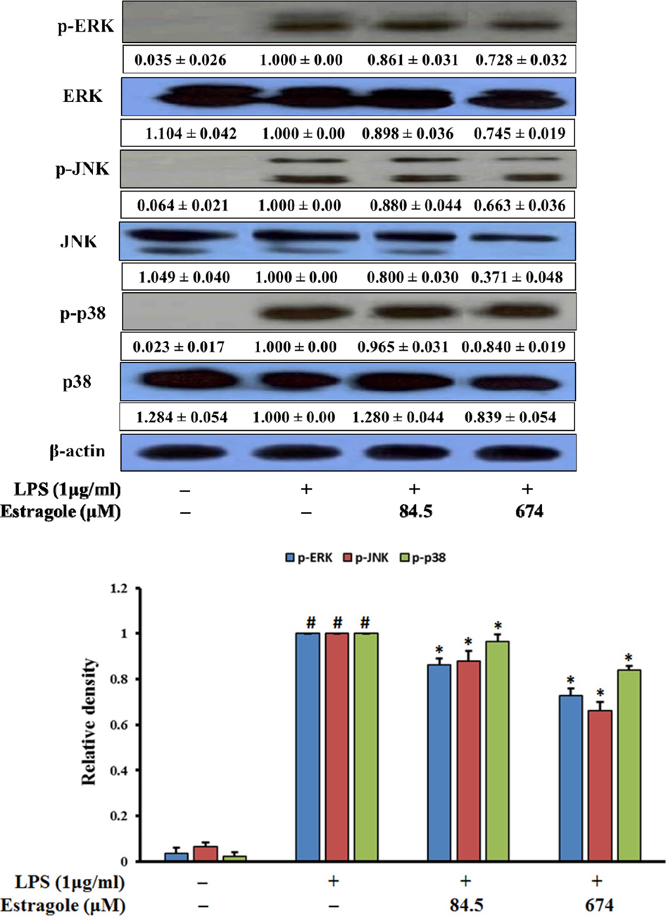
Fig. 4. Inhibitory effect of estragole on the expression of MAPKs in LPS-stimulated RAW264.7 cells. Cells were pretreated with the indicated concentrations of estragole for 2 h and stimulated with LPS (1 μg/mL) for 18 h. Data was detected by Western blot analysis with the designated antibodies. β-Actin was used as an internal control. The results presented are representative of three independent experiments. #p <0.05 indicates significant differences from the control group. ∗p < 0.05 indicates significant differences from the LPS-treated group.
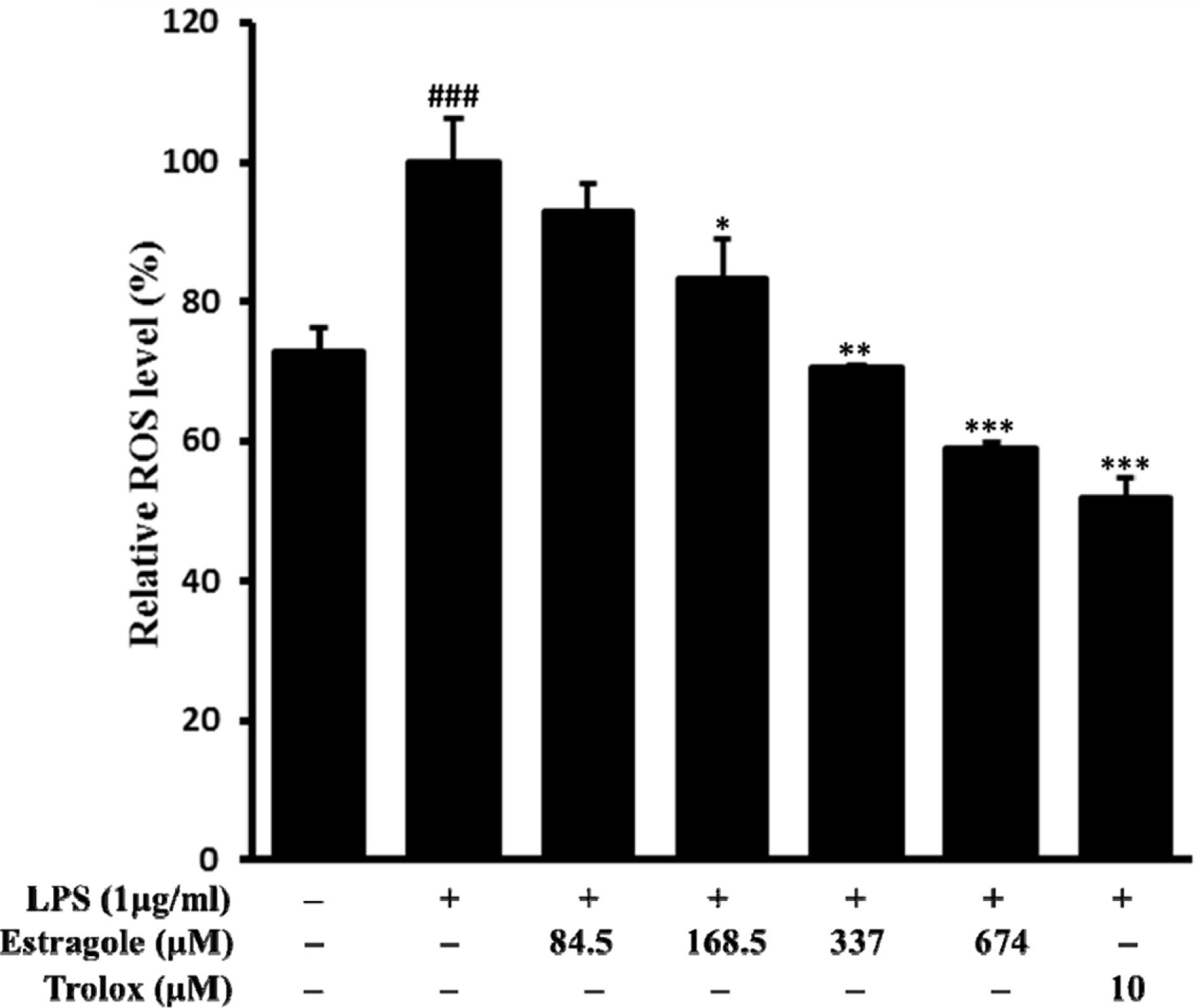
Fig. 5. Inhibitory effect of estragole on the production of ROS in LPS-stimulated RAW264.7 cells. Cells pretreated with different concentrations of estragole for 2 h were stimulated with LPS (1 μg/mL) for 24 h. ROS levels were measured by DCFH-DA with fluorescent analysis. Data are presented as the mean ± standard deviation of three independent experiments. ###p < 0.01 indicates significant differences from the control group. ∗p < 0.05, ∗∗p <0.01, and ∗∗∗p <0.001 indicate significant differences from the LPS-treated group.

Fig. 6. Inhibitory effect of estragole on the expression of HO-1 and Nrf-2 in LPS-stimulated RAW264.7 cells. Cells were pretreated with the indicated concentration of estragole for 2 h and stimulated with LPS (1 μg/mL) for 18 h. Data was detected by Western blot analysis with the designated antibodies. β-Actin was used as an internal control. The results presented were representative of three independent experiments. #p < 0.05 indicates significant differences from the control group. ∗p < 0.05 indicates significant differences from the LPS-treated group.
Reference
-
References
(1). Joung E. -J.., Lee B.., Gwon W. -G.., Shin T.., Jung B. -M.., Yoon N. -Y.., Choi J. S.., Oh C. -W.., Kim H.-R. Int. Immunopharmacol. 2005. 29:693–700.(2). Laroux F. S.., Pavlick K. P.., Hines I. N.., Kawachi S.., Harada H.., Bharwani S.., Hoffman J. M.., Grisham M. B.Acta Physiol. Scand. 2001. 173:113–118.(3). Giuliani C.., Napolitano G.., Bucci I.., Montani V.., Monaco F.Clin. Ter. 2001. 152:249–253.(4). May M. J.., Ghosh S.Immunol. Today. 1998. 19:80–88.(5). Tak P. P.., Firestein G. S. J.Clin. Invest. 2001. 107:7–11.(6). Cargnello M.., Roux P. P.Microbiol. Mol. Biol. Rev. 2011. 75:50–83.(7). Chen H. -G.., Xie K. -L.., Han H. -Z.., Wang W. -N.., Liu D. -Q.., Wang G. -L.., Yu Y. -H.Int. J. Surg. 2013. 11:1060–1066.(8). Taha R.., Blaise G.Funct. Food Health Dis. 2014. 4:510–523.(9). Bakkali F.., Averbeck S.., Averbeck D.., Idaomar M.Food Chem. Toxicol. 2008. 46:446–475.(10). Choi J. S.., Song B. M.., Park H. J.Kor. J. Pharmacogn. 2016. 47:192–196.(11). Donati M.., Mondin A.., Chen Z.., Miranda F. M.., do Nascimento B. B. Jr.., Schirato G.., Pastore P.., Froldi G.Nat. Prod. Res. 2015. 29:939–946.(12). Friedman M.., Henika P. R.., Mandrell R. E. J.Food Prot. 2002. 65:1545–1560.(13). Cosentino R. M.., Norte M. C.., Lazarini C. A.Phytother. Res. 2004. 18:921–924.(14). Albuquerque A. A.., Sorenson A. L.., Leal-Cardoso J. H. J.Ethnopharmacol. 1995. 49:41–49.(15). Ponte E. L.., Sousa P. L.., Rocha M. V.., Soares P. M.., Coelho-de-Souza A. N.., Leal-Cardoso J. H.., Assreuy A. M.Pharmacol. Rep. 2012. 64:984–990.(16). Rodrigues L. B.., Oliveira Brito Pereira Bezerra Martins A.., Cesário F. R.., Ferreira e Castro F.., de Albuquerque T. R.., Martins Fernandes M. N.., Fernandes da Silva B. A.., Quintans Júnior L. J.., da Costa J. G.., Melo Coutinho H. D.., Barbosa R.., Alencar de Menezes I. R.Chem. Biol. Interact. 2016. 257:14–25.(17). Keiser K.., Johnson C. C.., Tipton D. A. J.Endod. 2000. 26:288–291.(18). Meda L.., Cassatella M. A.., Szendrei G. I.., Otvos L. Jr.., Baron P.., Villalba M.., Ferrari D.., Rossi F.Nature. 1995. 374:647–650.(19). Dandona P.., Chaudhuri A.., Dhindsa S.Diabetes Care. 2010. 33:1686–1687.(20). Marks-Konczalik J.., Chu S. C.., Moss J. J.Biol. Chem. 1998. 273:22201–22208.(21). Islam M. N.., Choi R. J.., Jin S. E.., Kim Y. S.., Ahn B. R.., Zhao D.., Jung H. A.., Choi J. S. J.Ethnopharmacol. 2012. 144:175–181.(22). Chen J. -J.., Huang W. -C.., Chen C.-C. Mol. Biol. Cell. 2005. 16:5579–5591.(23). Rajapakse N.., Kim M. M.., Mendis E.., Kim S. K.Immunology. 2008. 123:348–357.(24). Kaminska B.Biochem. Biophys. Acta. 2005. 1754:253–262.(25). Xia Z.., Dickens M.., Raingeaud J.., Davis R. J.., Greenberg M. E.Science. 1995. 24:1326–1331.(26). Hancock J. T.., Desikan R.., Neill S. J.Biochem. Soc. Trans. 2001. 29:345–350.(27). Choi S. Y.., Hwang J. H.., Ko H. C.., Park J. G.., Kim S. J. J.Ethnopharmacol. 2007. 113:149–155.(28). Siomek A.Acta Biochem. Pol. 2012. 59:323–331.(29). Ryan K. A.., Simth M. F. Jr.., Sanders M. K.., Ernst P. B.Infect. Immun. 2004. 72:2123–2130.(30). Kim J. -H.., Choo Y. -Y.., Tae N.., Min B. -S.., Lee J.-H. Int. Immunopharmacol. 2014. 22:420–426.(31). Lee I. -S.., Lim J.., Gal J.., Kang J. -C.., Kim H. -J.., Kang B. -Y.., Choi H.-J. Neurochem. Int. 2011. 58:153–160.(32). Paine A.., Eiz-Vesper B.., Blasczyk R.., Immenschuh S.Biochem. Pharmacol. 2010. 80:1895–1903.(33). Lee M. -Y.., Lee J. -A.., Seo C.-S.., Ha H.., Lee H.., Son J. -K.., Shin H.-K. Food Chem. Toxicol. 2011. 49:1047–1055.(34). Tsoyi K.., Lee T. Y.., Lee Y. S.., Kim H. J.., Seo H. G.., Lee J. H.., Chang K. C.Mol. Pharmacol. 2009. 76:173–182.
- Full Text Links
-
- Actions
-
Cited
- CITED
-
- Close
- Share
-
- Similar articles
-
- Pulegone Exhibits Anti-inflammatory Activities through the Regulation of NF-κB and Nrf-2 Signaling Pathways in LPS-stimulated RAW 264.7 cells
- Luteolin 5-O-glucoside from Korean Milk Thistle, Cirsium maackii, Exhibits Anti-Inflammatory Activity via Activation of the Nrf2/HO-1 Pathway
- Anti-inflammatory activities of Scolopendra subspinipes mutilans in RAW 264.7 cells
- Aromadendrin Inhibits Lipopolysaccharide-Induced Nuclear Translocation of NF-kappaB and Phosphorylation of JNK in RAW 264.7 Macrophage Cells
- Anti-inflammatory effects of proanthocyanidin-rich red rice extract via suppression of MAPK, AP-1 and NF-κB pathways in Raw 264.7 macrophages
