Pediatr Gastroenterol Hepatol Nutr.
2018 Apr;21(2):86-92. 10.5223/pghn.2018.21.2.86.
Comparison of Endoscopic and Histological Findings between Typical and Atypical Celiac Disease in Children
- Affiliations
-
- 1Department of Paediatric Medicine, Sawai Man Singh Medical College, Jaipur, India. rkguptadr@hotmail.com
- 2Department of Community Medicine, University College of Medical Sciences, Delhi, India.
- KMID: 2409359
- DOI: http://doi.org/10.5223/pghn.2018.21.2.86
Abstract
- PURPOSE
Celiac disease is a common non-communicable disease with varied presentations. Purpose of this study was to find the duodeno-endoscopic features in celiac disease and to compare duodeno-endoscopic and histological findings between typical and atypical celiac disease in children.
METHODS
Hospital based observational study was conducted at Sir Padampat Mother and Child Health Institute, Jaipur from June 2015 to May 2016. Patients were selected and divided in two groups- typical and atypical celiac disease based upon the presenting symptoms. Upper gastrointestinal endoscopy and duodenal biopsy was performed for serology positive patients. Results were analysed using appropriate statistical test of significance.
RESULTS
Out of 101 enrolled patients, 47.5% were male. Age ranged from 1 to 18 years. Study showed that 54.5% were typical and 45.5% were atypical. Patients presenting with atypical symptoms were predominantly of older age group. On endoscopy, scalloping, mosaic pattern, reduced fold height and absent fold height; and in histology, advanced Marsh stage were significantly higher in the typical group.
CONCLUSION
Awareness of atypical presentations as well as duodeno-endoscopic features may have considerable practical importance for the diagnosis of celiac disease in children. Scalloping, mosaic pattern, reduced fold height and nodularity are main endoscopic markers of celiac disease in children. Endoscopic markers of duodenal mucosa may be important in early diagnosis of celiac disease, in children subjected to endoscopy for atypical presentations or indication other than suspected celiac disease.
Keyword
MeSH Terms
Figure
-
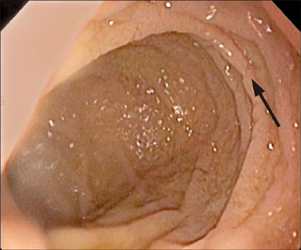
Fig. 1 Duodeno-endoscopic finding (arrow): scalloping.

Fig. 2 Duodeno-endoscopic finding (arrow): mosaic pattern.

Fig. 3 Duodeno-endoscopic finding (arrow): nodularity.
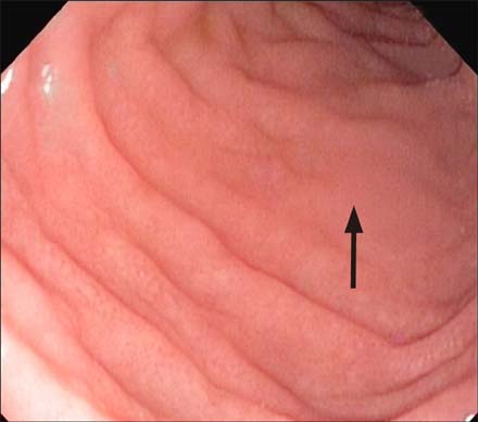
Fig. 4 Duodeno-endoscopic finding (arrow): reduced fold height.
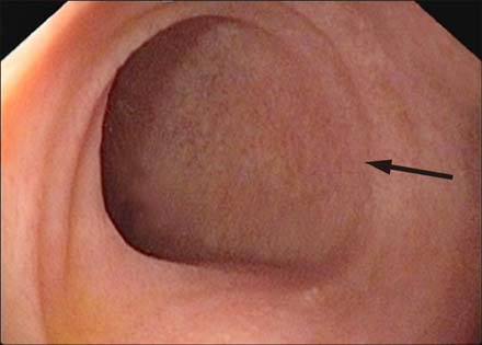
Fig. 5 Duodeno-endoscopic finding (arrow): absent fold height.
Reference
-
1. Husby S, Koletzko S, Korponay-Szabó IR, Mearin ML, Phillips A, Shamir R, et al. European Society for Pediatric Gastroenterology, Hepatology, and Nutrition guidelines for the diagnosis of coeliac disease. J Pediatr Gastroenterol Nutr. 2012; 54:136–160.
Article2. Lionetti E, Catassi C. New clues in celiac disease epidemiology, pathogenesis, clinical manifestations, and treatment. Int Rev Immunol. 2011; 30:219–231.
Article3. Branski D, Troncone R. Gluten sensitive enteropathy (celiac disease). In : Kliegman RM, Stanton BF, St Geme JW, Schor NF, Behrman RE, Nelson WE, editors. Nelson textbook of pediatrics. 20th ed. Philadelphia: Elsevier Saunders;2016. p. 1835–1838.4. Dinler G, Atalay E, Kalayci AG. Celiac disease in 87 children with typical and atypical symptoms in Black Sea region of Turkey. World J Pediatr. 2009; 5:282–286.
Article5. Guandalini S. Celiac disease. In : Guandalini S, editor. Textbook of pediatric gastroenterology and nutrition. London: Taylor & Francis;2004. p. 435–450.6. Sharma A, Poddar U, Yachha SK. Time to recognize atypical celiac disease in Indian children. Indian J Gastroenterol. 2007; 26:269–273.7. Garg K, Gupta RK. What a practitioner needs to know about celiac disease? Indian J Pediatr. 2015; 82:145–151.
Article8. Balaban DV, Popp A, Vasilescu F, Haidautu D, Purcarea RM, Jinga M. Diagnostic yield of endoscopic markers for celiac disease. J Med Life. 2015; 8:452–457.9. Ravelli AM, Tobanelli P, Minelli L, Villanacci V, Cestari R. Endoscopic features of celiac disease in children. Gastrointest Endosc. 2001; 54:736–742.
Article10. Rosa RM, Ferrari Mde L, Pedrosa MS, Ribeiro GM, Brasileiro-Filho G, Cunha AS. Correlation of endoscopic and histological features in adults with suspected celiac disease in a referral center of Minas Gerais, Brazil. Arq Gastroenterol. 2014; 51:290–296.
Article11. Corazza GR, Caletti GC, Lazzari R, Collina A, Brocchi E, Di Sario A, et al. Scalloped duodenal folds in childhood celiac disease. Gastrointest Endosc. 1993; 39:543–545.
Article12. Kasirer Y, Turner D, Lerman L, Schechter A, Waxman J, Dayan B, et al. Scalloping is a reliable endoscopic marker for celiac disease. Dig Endosc. 2014; 26:232–235.
Article13. Oberhuber G, Granditsch G, Vogelsang H. The histopathology of coeliac disease: time for a standardized report scheme for pathologists. Eur J Gastroenterol Hepatol. 1999; 11:1185–1194.14. Poddar U. Pediatric and adult celiac disease: similarities and differences. Indian J Gastroenterol. 2013; 32:283–288.
Article15. Gupta R, Reddy DN, Makharia GK, Sood A, Ramakrishna BS, Yachha SK, et al. Indian task force for celiac disease: current status. World J Gastroenterol. 2009; 15:6028–6033.
Article16. Kuloğlu Z, Kirsaçlioğlu CT, Kansu A, Ensari A, Girgin N. Celiac disease: presentation of 109 children. Yonsei Med J. 2009; 50:617–623.
Article17. Savas N, Akbulut S, Saritas U, Koseoglu T. Correlation of clinical and histopathological with endoscopic findings of celiac disease in the Turkish population. Dig Dis Sci. 2007; 52:1299–1303.
Article18. Piazzi L, Zancanella L, Chilovi F, Merighi A, De Vitis I, Feliciangeli G, et al. Diagnostic value of endoscopic markers for celiac disease in adults: a multicentre prospective Italian study. Minerva Gastroenterol Dietol. 2008; 54:335–346.19. Tursi A, Brandimarte G, Giorgetti GM, Gigliobianco A. Endoscopic features of celiac disease in adults and their correlation with age, histological damage, and clinical form of the disease. Endoscopy. 2002; 34:787–792.
Article20. Brar P, Kwon GY, Egbuna II, Holleran S, Ramakrishnan R, Bhagat G, et al. Lack of correlation of degree of villous atrophy with severity of clinical presentation of coeliac disease. Dig Liver Dis. 2007; 39:26–29.
Article
- Full Text Links
-
- Actions
-
Cited
- CITED
-
- Close
- Share
-
- Similar articles
-
- Association between Celiac Disease and Intussusceptions in Children: Two Case Reports and Literature Review
- Atypical Symptoms in Patients With Gastroesophageal Reflux Disease
- Clinical Analysis of Atypical Kawasaki Disease: Comparison of Kawasaki Disease Between Typical and Atypical Types
- Lobar Atelectasis: Typical and Atypical Radiographic and CT Findings
- Seronegative Celiac Disease in Patients with Isolated Refractory Dyspepsia and Gastroesophageal Reflux Disease
