Perinatology.
2017 Dec;28(4):162-165. 10.14734/PN.2017.28.4.162.
Miller-Dieker Syndrome in an Extremely Low Birth Weight Infant
- Affiliations
-
- 1Department of Pediatrics, Pusan National University School of Medicine, Busan, Korea. kuma2002@hanmail.net
- KMID: 2409125
- DOI: http://doi.org/10.14734/PN.2017.28.4.162
Abstract
- Miller-Dieker syndrome (MDS) is characterized by severe lissencephaly and facial dysmorphism including the prominent forehead, bitemporal hollowing, a short nose with upturned nares, a protuberant upper lip, and a small jaw. MDS can be caused by deletions or mutations of the LIS1 gene on 17p13.3. Our patient was born at 28⺳ weeks gestation and weight 950 g at birth. He was suffered from respiratory distress syndrome and feeding intolerance. And he showed colpocephaly with agenesis of corpus callosum on the first brain ultrasonography. So, brain magnetic resonance imaging was performed 2 months of age and revealed agyria, colpocephaly, agenesis of corpus callosum and band heterotopias. At initial physical examination revealed no specific abnormal findings. However, hypotonia and abnormal facial morphology including prominent forehead, a short nose with upturned nares and protuberant upper lip were developed gradually as he got older. Chromosomal microarray was performed and confirmed microdeletion on 17p13.3. In conclusion, MDS should be considered when the baby shows colpocephaly, agenesis of corpus callosum, hypotonia, feeding problem and abnormal facial features even though the baby is preterm infant.
MeSH Terms
-
Agenesis of Corpus Callosum
Brain
Classical Lissencephalies and Subcortical Band Heterotopias*
Forehead
Humans
Infant
Infant, Extremely Low Birth Weight*
Infant, Newborn
Infant, Premature
Infant, Very Low Birth Weight
Jaw
Lip
Lissencephaly
Magnetic Resonance Imaging
Muscle Hypotonia
Nose
Parturition
Physical Examination
Pregnancy
Ultrasonography
Figure
-
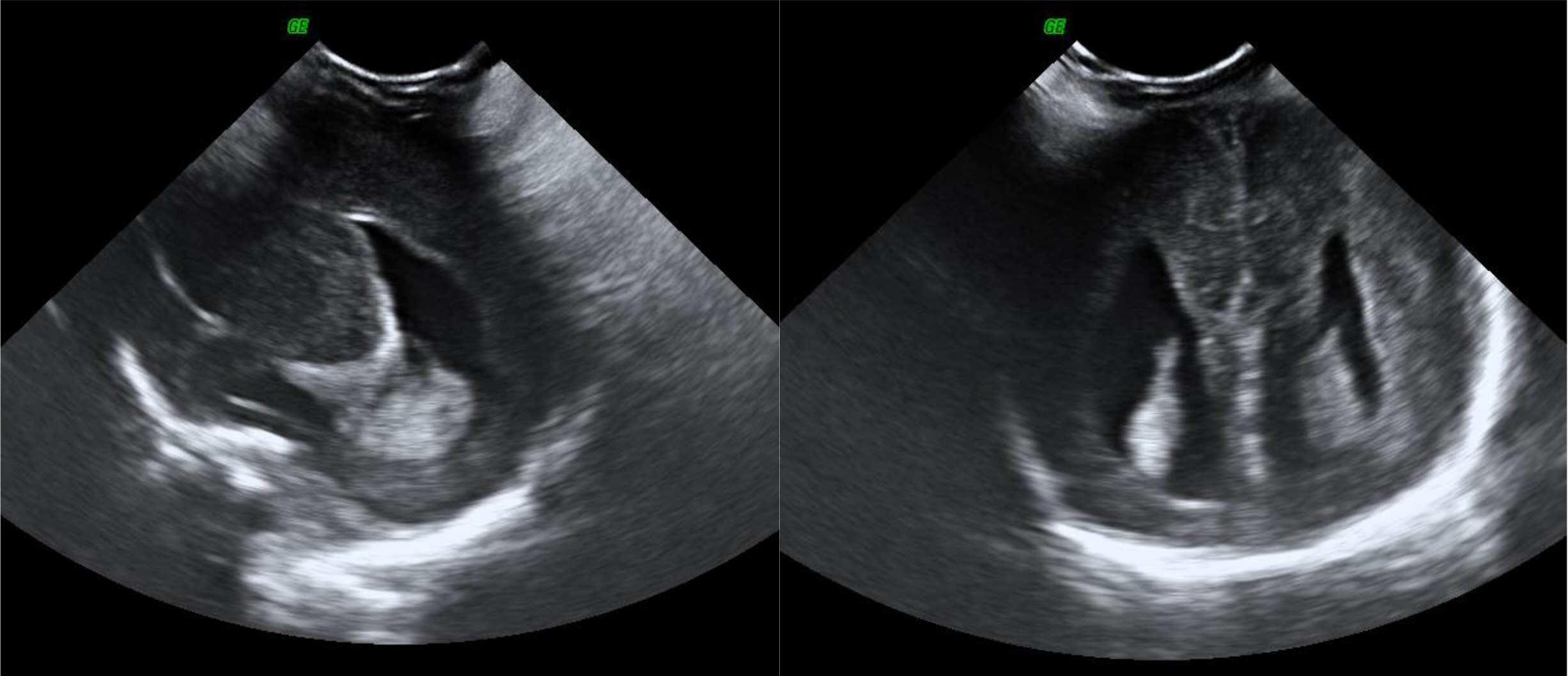
Fig. 1 The initial brain ultrasonography showed colpocephaly and agenesis of corpus callosum at 3 days of age.
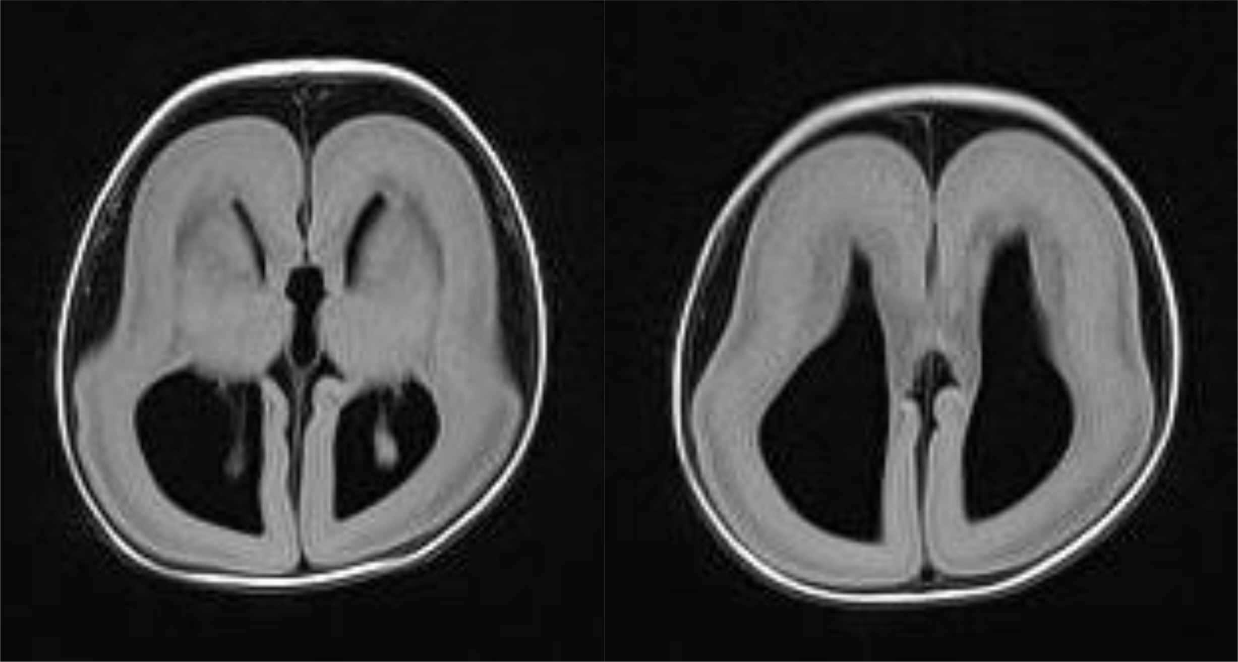
Fig. 2 A brain magnetic resonance imaging showed agyria, colpocephaly, agenesis of corpus callosum and band heterotopia at 2 months of age.

Fig. 3 The patient showed prominent forehead, a short nose with upturned nares and a protuberant upper lip.
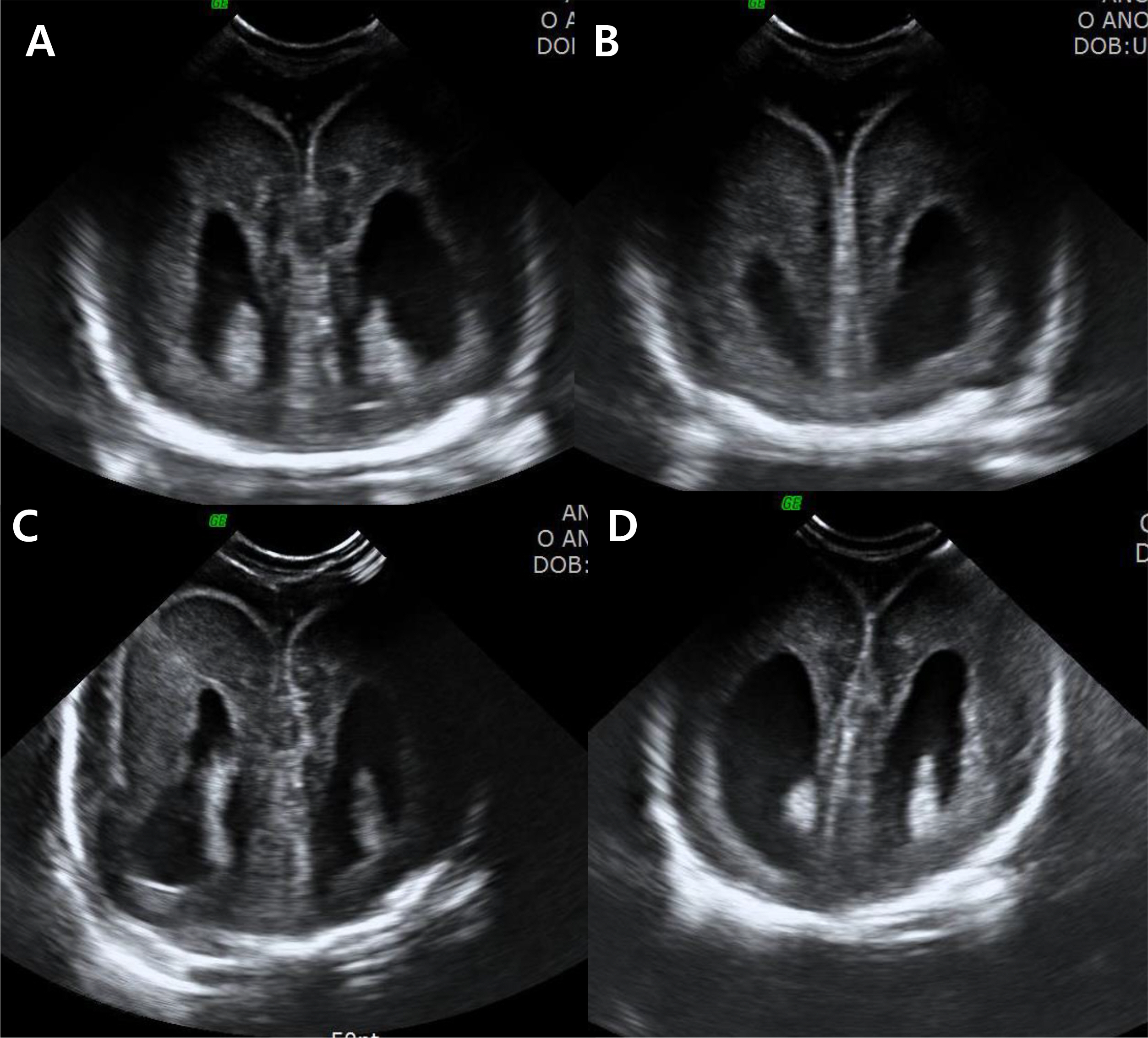
Fig. 4 The serial brain ultrasonography showed agyria at (A) 4 weeks, (B) 5 weeks, (C) 6 weeks, and (D) 8 weeks of age respectively.
Reference
-
1). Dobyns WB., Curry CJ., Hoyme HE., Turlington L., Ledbetter DH. Clinical and molecular diagnosis of Miller-Dieker syndrome. Am J Hum Genet. 1991. 48:584–94.2). Reiner O., Carrozzo R., Shen Y., Wehnert M., Faustinella F., Dobyns WB, et al. Isolation of a Miller-Dieker lissencephaly gene containing G protein beta-subunit-like repeats. Nature. 1993. 364:717–21.3). Monteagudo A., Timor-Tritsch IE. Development of fetal gyri, sulci and fissures: a transvaginal sonographic study. Ultrasound Obstet Gynecol. 1977. 9:222–8.
Article4). Miller JQ. Lissencephaly in 2 siblings. Neurology. 1963. 13:841.
Article5). Garcia CA., Dunn D., Trevor R. The lissencephaly (agyria) syndrome in siblings. Arch Neurol. 1978. 35:608–11.
Article6). Daube JR., Chou SM. Lissencephaly: two case. Neurology. 1966. 16:179–91.7). Greenberg F., Courtney KB., Wessels RA., Huhta J., Carpenter RJ., Rich DC, et al. Prenatal diagnosis of deletion 17p13 associated with DiGeorge anomaly. Am J Med Genet. 1988. 31:1–4.
Article8). Lin CY., Chen CP., Liau CL., Su PH., Tsao TF., Chang TY, et al. Prenatal diagnosis of monosomy 17p (17p13.3→pter) associated with polyhydramnios, intrauterine growth restriction, ventriculomegaly, and Miller-Dieker lissencephaly syndrome in a fetus. Taiwan J Obstet Gynecol. 2009. 48:408–11.9). McGahan JP., Grix A., Gerscovich EO. Prenatal diagnosis of lissencephaly: Miller-Dieker syndrome. J Clin Ultrasound. 1994. 22:560–3.
Article10). Chitayat D., Toi A., Babul R., Blaser S., Moola S., Yarkoni D, et al. Omphalocele in Miller-Dieker syndrome: expanding the phenotype. Am J Med Genet. 1997. 69:293–8.
Article11). Lenzini E., D'Ottavio G., Città A., Benussi DG., Petix V., Peoile V. Perinatal diagnosis of Miller-Dieker syndrome by ultrasound and molecular cytogenetic analysis. Clin Genet. 2007. 72:487–9.12). Guerrini R., Filippi T. Neuronal migration disorders, genetics, and epilep-togenesis. J Child Neurol. 2005. 20:287–99.
- Full Text Links
-
- Actions
-
Cited
- CITED
-
- Close
- Share
-
- Similar articles
-
- A Case of Miller-Dieker Syndrome with Infantile Spasm and Lennox-Gastaut Syndrome
- Successful Peritoneal Dialysis in an Extremely Preterm Infant
- Meconium Obstruction Syndrome in Two Extremely Low Birth Weight Infants
- Early-Onset Sepsis Due to Listeria Monocytogenes in a Extremely Low Birth Weight Infant
- Socioeconomic Disparities in Pregnancy Outcome and Infant Mortality: Extremely Low Birth Weight and Very Low Birth Weight Infants
