Successful Treatment of a Term Neonate Developing Severe Pulmonary Interstitial Emphysema Unrelated to Mechanical Ventilation; the First Reported Case in Korea
- Affiliations
-
- 1Department of Pediatrics, Kangwon National University Hospital, Chuncheon, Korea. sysmile@gmail.com
- KMID: 2409114
- DOI: http://doi.org/10.14734/PN.2018.29.1.57
Abstract
- Air leak syndrome is one of the main causes of respiratory failure which is defined as air escapes from intra-alveolar to extra-alveolar areas. The incidence of spontaneous air leak syndrome in premature infants is about 1-2%. Pulmonary interstitial emphysema (PIE) is a form of air leak syndrome which usually occurs in infants with poor lung compliance (such as respiratory distress syndrome) on inadequate mechanical ventilation. On the other hand, air leak syndrome in term infants without underlying lung disease is very rare. Especially, there is only one report in Korea regarding PIE in term infants who finally underwent surgery. We experienced a case of a term infant who developed severe PIE unrelated to conventional mechanical ventilation and successfully treated by high-frequency ventilation care without any invasive procedure.
MeSH Terms
Figure
-

Fig. 1 Infantograms of the patient during admission. No abnormality at admission (A) and on CPAP (B). After respiratory deterioration, infantogram just after intubation (C) showed pneumomediastinum, small amount of right pneumothorax, and, PIE (arrow) at right lung. Resolved air leak after HFV (D). CPAP, continuous positive airway pressure; PIE, pulmonary interstitial emphysema; HFV, high frequency ventilation.
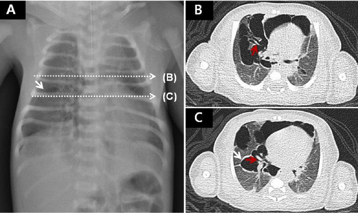
Fig. 2 Chest CT images (B, C) with correlation of the infantogram (A). White arrows (A, C) indicate RML PIE which is visible at the infantogram. Venules and arterioles (red arrows [B, C]) normally located inside bronchovascular sheath, are surrounded by huge amount of air suggesting PIE in the right lung. Chest CT also shows air extending to the mediastinum and pleural space (B, C). CT, computed tormography; RML, right middle lung; PIE, pulmonary interstitial emphysema.
Reference
-
1. Bawa P, Soontarapornchai K, Perenyi A, Goldfisher R, Amodio J. Development of Localized Pulmonary Interstitial emphysema in a late preterm infant without mechanical ventilation. Case Rep Pediatr. 2014; 2014:429797.
Article2. Freysdottir D, Olutoye O, Langston C, Fernandes CJ, Tatevian N. Spontaneous pulmonary interstitial emphysema in a term unventilated infant. Pediatr Pulmonol. 2006; 41:374–378.
Article3. Macklin MT, Macklin CC. Malignant interstitial emphysema of the lungs and mediastinum as an important occult complication in many respiratory diseases and other conditions: an interpretation of the clinical literature in the light of laboratory experiment. Medicine. 1944; 23:281–358.4. Greenough A, Dixon AK, Roberton NR. Pulmonary interstitial emphysema. Arch Dis Child. 1984; 59:1046–1051.
Article5. Jeng MJ, Lee YS, Tsao PC, Soong WJ. Neonatal air leak syndrome and the role of high-frequency ventilation in its prevention. J Chin Med Assoc. 2012; 75:551–559.
Article6. Rent SM, Donn SM. Treatment of severe unilateral pulmonary interstitial emphysema in a preterm infant. Ann Med Case Rep. 2017; 1:1019.7. Morisot C, Kacet N, Bouchez MC, Rouland V, Dubos JP, Gremillet C, et al. Risk factors for fatal pulmonary interstitial emphysema in neonates. Eur J Pediatr. 1990; 149:493–495.
Article8. Aiyoshi T, Masumoto K, Shinkai T, Tanaka Y, Fujii S, Sasaki T, et al. Pulmonary interstitial emphysema due to respiratory syncytial virus infection. Pediatr Int. 2016; 58:916–919.
Article9. Morley CJ, Davis PG, Doyle LW, Brion LP, Hascoet JM, Carlin JB. Nasal CPAP or intubation at birth for very preterm infant. N Engl J Med. 2008; 358:700–708.10. Bhojani S, Bird D, Alok G. Spontaneous diffuse pulmonary interstitial emphysema (PIE) in an unventilated infant. Internet J Pediatr Neonatol. 2008; 9:2.
Article11. Clark RH, Gerstmann DR, Null DM, Yoder BA, Cornish JD, Glasier CM, et al. Pulmonary interstitial emphysema treated by high-frequency oscillatory ventilation. Crit Care Med. 1986; 14:926–930.
Article12. Keszler M, Modanlou HD, Brudno DS, Clark FI, Cohen RS, Ryan RM, et al. Multicenter controlled clinical trial of high-frequency jet ventilation in preterm infants with uncomplicated respiratory distress syndrome. Pediatrics. 1997; 100:593–599.
Article13. Jassal MS, Benson JE, Mogayzel PJ Jr. Spontaneous resolution of diffuse persistent pulmonary interstitial emphysema. Pediatric Pulmonol. 2008; 43:615–619.
Article14. Oh MH, Kim MY, Shim WS, Oh SS, Shin BK, Cho SJ, et al. A case of localized persistent interstitial pulmonary emphysema. J Korean Med Sci. 2001; 16:225–228.
Article
- Full Text Links
-
- Actions
-
Cited
- CITED
-
- Close
- Share
-
- Similar articles
-
- A case of localized persistent interstitial pulmonary emphysema
- Development of pulmonary air leak in an extremely-low-birth-weight infant without mechanical ventilation: a case report
- A Case of high-Frequency Oscillatory Ventilation
- A Case of Acute Respiratory Distress Syndrome Occurred after Pulmonary Edema in a Fullterm Newborn
- A Case of Nonspecific Interstitial Pneumonia Complicated with Spontaneous Pneumomediastinum, Subcutaneous Emphysema and Pneumatosis Interstinalis
